Volume 13, Number 8—August 2007
Letter
Alistipes finegoldii in Blood Cultures from Colon Cancer Patients
Figure
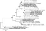
Figure. Phylogenetic tree inferred from comparison of the 16S rRNA gene sequences of genera Bacteroides, Parabacteroidetes, Prevotella, and Alistipes. Nucleotide accession numbers for the sequences used to construct this dendrogram are given...
To the Editor: Alistipes finegoldii was previously isolated from appendiceal tissue samples in children with acute appendicitis and from perirectal and brain abscess material (1,2). 16S rDNA sequencing studies showed that this bacterium clustered with A. putredinis (Figure) in the Bacteroidetes group (4). We describe the first cases, to our knowledge, of bacteremia due to A. finegoldii in 2 patients with colon cancer who underwent surgical resection.
The first patient was a 61-year-old woman with colorectal carcinoma and liver metastasis, who underwent chemotherapy consisting of 6 cycles of oxaliplatin (the FOLFOX scheme, a chemotherapy regimen consisting of fluorouracil [5 FU], folinic acid, and oxaliplatin). In September 2003, a left colectomy, resection of metastasis in the left side of the liver, and a ligation of the right portal vein were performed. Two months later, in a second step, a right hepatectomy was done. On postoperative day 5, the patient had a fever up to 39.8°C and leukocyte count of 8.49 g/L (68% polymorphonuclear leukocytes). Two blood cultures were performed before antimicrobial drug therapy based on amoxicillin/clavulanic acid and amikacin was started. After receiving this therapy, the patient recovered rapidly. One of the 2 anaerobic blood cultures was positive. Gram-negative bacilli were isolated (strain 3302398). Antimicrobial susceptibility testing showed decreased susceptibility to vancomycin, cefotetan, and penicillin G. The strain produced β-lactamase as determined by Cefinase test (Becton Dickinson, Le Pont de Claix, France).
The second patient was a 64-year-old man with colon cancer who was receiving palliative chemotherapy (16th cycle, FOLFOX scheme); he was seen in March 2004 with a fever up to 39°C. An adenocarcinoma of the ileum had been diagnosed in June 2002 in this patient, and an ileocecal resection was performed followed by adjuvant chemotherapy. One year later, a local recurrence and peritoneal carcinomatosis were detected. The patient again underwent abdominal surgery by resection of ileo-colic anastomosis and sigmoid and peritoneal masses; a colostomy had to be created. The patient’s leukocyte count was 14.94 g/L (84.6% polymorphonuclear leukocytes), and his C-reactive protein level was 268 mg/L. Before antimicrobial drug therapy with amoxicilline/clavulanic acid and ciprofloxacin was begun, blood cultures were taken. One of the 2 anaerobic blood cultures was positive. Gram-negative bacilli were isolated (strain 4401054). Antimicrobial drug resistance was detected only to vancomycin. After receiving this therapy, the patient recovered rapidly.
Biochemical characterization was conducted by using API 20A and rapid ID 32A strips (bioMérieux, Marcy l’Etoile, France). Results were compared with those obtained for the reference strain A. finegoldii CIP 107999T. Strains 3302398 and 4401054 were indole positive and bile resistant, and they had positive enzyme reactions for N-acetyl-β-glucosaminidase, α-galactosidase, and β-galactosidase, as described for A. finegoldii (4). The 2 strains produced a brown pigment after 2 weeks’ incubation on sheep blood agar plates (bioMérieux).
PCR amplification of the 16S rDNA was performed with the primer pair fD1/rp2 (5). The generated fragments were sequenced as previously described (6). Sequences were compared with those available in GenBank databases by using BLAST (www.ncbi.nlm.nih.gov/blast). They showed a 97% identity to the 16S rDNA of A. finegoldii (accession nos. AY643083 and AY643084).
A novel bacterium was characterized from appendiceal tissues samples from children with appendicitis and in 2 cases of perirectal and brain abscesses associated with other anaerobes (1). With routine tests, this organism resembled members of the Bacteroides fragilis group; however, the cellular fatty acid composition dominated by iso-C15:0 and production of brown pigment on media containing hemolyzed blood suggested that the organism was most closely related to the genus Porphyromonas (1). However, 16S rDNA sequence comparison showed highest sequence relatedness with B. putredinis, and the reclassification of B. putredinis in a novel genus, Alistipes, and the classification of the novel bacterium as A. finegoldii were proposed (4). A. putredinis was characterized in the indigenous flora of the human gut (7). The natural habitat of A. finegoldii is unknown but is probably the same. B. fragilis is the most frequent anaerobic bacterium isolated from blood samples, and the principal source of the bacteria is the gastrointestinal tract (8). Predisposing factors to Bacteroides species bacteremia include malignant neoplasms, recent gastrointestinal or obstetric-gynecologic surgery, intestinal obstruction, and use of cytotoxic agents or corticosteroids (8). In both of our patients, fever was noted and no other microorganisms were isolated, indications that the bacteria probably were pathogenic.
Phenotypic identification of Alistipes sp. is difficult in a routine microbiology laboratory. However, a molecular approach based on 16S rRNA gene sequence comparison is a good method for identifying anaerobic bacteria, as it has recently been reported for B. fragilis in anaerobic sepsis (9) and for B. thetaiotaomicron from a patient with a cholesteatoma and purulent meningitis (10). In our 2 patients, we also used molecular identification because A. finegoldii was not included in the API phenotypic database identification. A. finegoldii should be considered as an agent of bacteremia in patients with gastrointestinal pathologic conditions.
Acknowledgment
This work was supported by grant PBBSB-102600 from the Swiss National Science Foundation.
References
- Rautio M, Lonnroth M, Saxen H, Nikku R, Väisanen ML, Finegold SM, Characteristics of an unusual anaerobic pigmented gram-negative rod isolated from normal and inflamed appendices. Clin Infect Dis. 1997;25(Suppl 2):S107–10. DOIPubMedGoogle Scholar
- Rautio M, Saxen H, Siitonen A, Nikku R, Jousimies-Somer H. Bacteriology of histopathologically defined appendicitis in children. Pediatr Infect Dis J. 2000;19:1078–83. DOIPubMedGoogle Scholar
- Kimura M. A simple method for estimating evolutionary rates of base substitutions through comparative studies of nucleotide sequences. J Mol Evol. 1980;16:111–20. DOIPubMedGoogle Scholar
- Rautio M, Eerola E, Väisänen-Tunkelrott ML, Molitoris D, Lawson P, Collins MD, Reclassification of Bacteroides putredinis (Weinberg et al., 1937) in a new genus Alistipes gen. nov., as Alistipes putredinis comb.nov., and description of Alistipes finegoldii sp. nov., from human sources. Syst Appl Microbiol. 2003;26:182–8. DOIPubMedGoogle Scholar
- Weisburg WG, Barns SM, Pelletier DA, Lane DJ. 16S ribosomal DNA amplification for phylogenetic study. J Bacteriol. 1991;173:697–703.PubMedGoogle Scholar
- Fenner L, Roux V, Mallet MN, Raoult D. Bacteroides massiliensis sp. nov., isolated from blood culture of a newborn. Int J Syst Evol Microbiol. 2005;55:1335–7. DOIPubMedGoogle Scholar
- Rigottier-Gois L, Rochet V, Garrec N, Suau A, Doré J. Enumeration of Bacteroides species in human faeces by fluorescent in situ hybridisation combined with flow cytometry using 16S rRNA probes. Syst Appl Microbiol. 2003;26:110–8. DOIPubMedGoogle Scholar
- Brook I. Clinical review: bacteremia caused by anaerobic bacteria in children. Crit Care. 2002;6:205–11. DOIPubMedGoogle Scholar
- Wareham DW, Wilks M, Ahmed D, Brazier JS, Millar M. Anaerobic sepsis due to multidrug-resistant Bacteroides fragilis: microbiological cure and clinical response with linezolid therapy. Clin Infect Dis. 2005;40:67–8. DOIPubMedGoogle Scholar
- Feuillet L, Carvajal J, Sudre I, Pelletier J, Thomassin JM, Drancourt M, First isolation of Bacteroides thetaiotaomicron from a patient with a cholesteatoma and experiencing meningitis. J Clin Microbiol. 2005;43:1467–9. DOIPubMedGoogle Scholar
Figure
Cite This ArticleRelated Links
Table of Contents – Volume 13, Number 8—August 2007
| EID Search Options |
|---|
|
|
|
|
|
|
Please use the form below to submit correspondence to the authors or contact them at the following address:
Didier Raoult, Hôpital de la Timone, 264 rue Saint-Pierre, 13385 Marseille, France;
Top