Volume 14, Number 2—February 2008
Letter
Saksenaea vasiformis Infection, French Guiana
Denis Blanchet* , Eric Dannaoui†‡, Angela Fior*, Florence Huber*, Pierre Couppié*, Nour Salhab*, Damien Hoinard†, and Christine Aznar*
Figure
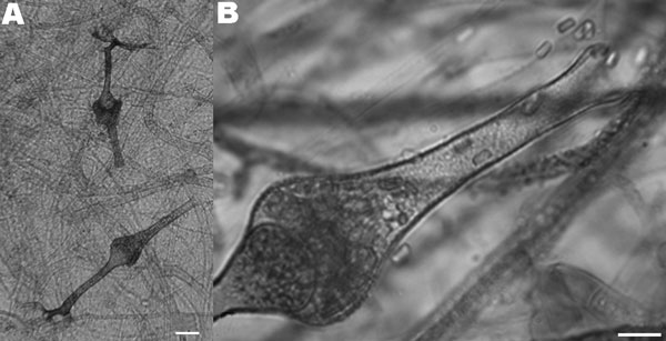
Figure. Microscopic characteristics of the isolate of Saksenaea vasiformis cultured on Czapek agar. A) Typical flask-shaped sporangia (scale bar = 25 μm) containing B) smooth-walled, rectangular sporangiospores (scale bar = 10 μm).
Page created: July 12, 2010
Page updated: July 12, 2010
Page reviewed: July 12, 2010
The conclusions, findings, and opinions expressed by authors contributing to this journal do not necessarily reflect the official position of the U.S. Department of Health and Human Services, the Public Health Service, the Centers for Disease Control and Prevention, or the authors' affiliated institutions. Use of trade names is for identification only and does not imply endorsement by any of the groups named above.