Volume 16, Number 2—February 2010
Dispatch
Increasing Incidence of Nontuberculous Mycobacteria, Taiwan, 2000–2008
Abstract
To assess the species distribution and epidemiologic trends of nontuberculous mycobacteria, we examined isolates from patients in Taiwan. During 2000–2008, the proportion increased significantly from 32.3% to 49.8%. Associated disease incidence increased from 2.7 to 10.2 cases per 100,000 patients. Mycobacterium avium complex and M. abscessus were most frequently isolated.
In Taiwan, the incidence of tuberculosis (TB) remains high despite advances in the antimycobacterial therapy and implementation of well-known TB control measures (1). Therefore, patients with acid-fast bacilli (AFB)–positive specimens, especially respiratory samples, are generally presumed to be infected with Mycobacterium tuberculosis and are treated with antituberculosis agents and placed in isolation rooms. Increased isolation of nontuberculous mycobacteria (NTM) causing mycobacterial diseases (2,3) implies that more patients with AFB-positive samples have received inappropriate or unnecessary empirical antituberculous treatment.
To help clinicians determine whether to initiate antituberculous treatment, mycobacterial laboratories should provide percentages of NTM isolates among specimens with positive AFB. However, the number and species of NTM from clinical specimens are increasing in the clinical laboratory, and the distribution of NTM species varies by geographic area (4). We thus sought to assess the species distribution of NTM isolates from various clinical specimens and to elucidate the epidemiologic trends of NTM isolates and diseases over a 9-year period in Taiwan.
To assess prevalence of NTM isolates, we used the database of the National Taiwan University Hospital mycobacterial laboratory and prepared clinical specimens for mycobacteria cultures according to recommended guidelines (5). Before 2001, specimens were spread onto Lowenstein-Jensen slants and Middlebrook 7H11 medium (BBL; Becton, Dickinson and Company Diagnostic Instrument Systems, Sparks, MD, USA); since late 2001, they have been spread onto Lowenstein-Jensen slants and tested by the fluorometric BACTEC system (BACTEC Mycobacterium Growth Indicator Tube 960 system; Becton, Dickinson and Company). NTM were identified to the species level by using conventional biochemical methods. Some unidentified NTM species were further confirmed by sequencing of their 16S rRNA gene (1,464 bp) by using 2 primers (8FPL and 1492) as previously described (6,7). Diseases caused by NTM were defined according to previous description (4). Annual incidence and isolation ratio of TB and NTM over time were evaluated by the Cochran-Armitage test for trend. We considered p<0.05 to be significant. To define NTM disease incidence and prevalence, for patients who had isolates over successive years during the study period we included only the year in which they first appeared in the laboratory database.
Figure
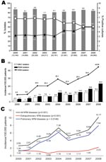
Figure. Incidence data for clinical samples submitted for mycobacterial culture, National Taiwan University Hospital, Taipei, Taiwan, January 2000–December 2008. A) Annual number and rate of isolates of nontuberculous mycobacteria (NTM) (triangles) and...
From January 2000 through December 2008, the laboratory received 283,394 clinical samples for mycobacterial culture. From a total of 23,499 (8.3%) specimens with positive mycobacterial culture results, M. tuberculosis were isolated from 14,295 (5.0%) specimens from 3,695 patients and NTM were isolated from 9,204 (3.2%) specimens from 4,786 patients. The trends of decreasing M. tuberculosis isolation and increasing NTM isolation were significant (p<0.05 for each) (Figure, panel A).
Among the 9,204 NTM isolates, M. avium complex (MAC, 30.0%) were the most frequently isolated organisms, followed by M. abscessus (17.5%), M. fortuitum complex (13.0%), M. chelonae complex (9.6%), M. kansasii (5.6%), and M. gordonae (5.5%) (Table). Among NTM isolates from the 4,786 patients, prevalent species were MAC (31.7%), M. fortuitum complex (18.2%), M. abscessus (17.2%), M. gordonae (11.6%), M. chelonae complex (8.2%), and M. kansasii (6.0%) (Table). In addition, some rare isolates such as M. celatum (n = 3), M conceptionene (n = 3), M. neoaurum (n = 2), M. arupense (n = 1), M. mageritense (n = 1), M. asiaticum (n = 1), and M. immunogenum (n = 1) were identified.
Annual incidences are shown in the Figure, panels B and C. During the study period, the proportions of NTM and rapidly growing mycobacteria among all mycobacteria isolated increased significantly from 32.3% to 49.8% (p<0.05) and from 3.7% to 23.4% (p<0.05), respectively. Incidence of diseases caused by NTM also increased (p<0.0001) from 2.7 to 10.2 per 100,000 inpatients and outpatients. The incidences of pulmonary and extrapulmonary NTM infection significantly increased from 1.26 to 7.94 per 100,000 inpatients (p<0.0001) and from 1.4 to 2.23 per 100,000 outpatients (p = 0.0194), and the increase of all NTM diseases was predominately in pulmonary diseases. During the 9 years, 1,105 patients had NTM diseases; most commonly pulmonary disease (n = 894, 76.8%), followed by soft tissue infection (n = 122, 11.4%), disseminated infection (n = 79, 7.1%), and peritonitis (n = 19, 1.7%). MAC was the most common cause of pulmonary infection (n = 342, 40.3%) and disseminated infection (n = 56, 70.1%); M. abscessus was the most common cause of skin and soft tissue infection (n = 46, 37.7%).
During the 9-year period, NTM accounted for 39.2% of positive mycobacterial cultures and increased significantly. In concordance with the increased incidence of NTM isolations, incidence of NTM diseases also increased significantly. The increase of NTM isolations since 2002 might have resulted from use of the BACTEC system. This apparent increase in NTM disease could also be attributed to increasing vigilance and awareness of these bacteria as human pathogens, improved methods of detection, or more immunocompromised hosts (e.g., as a result of tumor necrosis factor inhibitors, human interleukin 1 receptor antagonists, and anti-CD20 antibodies) (1,8).
Prevalence of mycobacteria species responsible for different diseases varies markedly by geographic region. In the United States and Japan, MAC and M. kansasii are the most common species (4), whereas in England and Scotland, M. kansasii and M. malmoense, respectively, are the most common (9). Our study showed that MAC was the most common NTM species in Taiwan, followed by M. abscessus. The most common organism in localized pulmonary infection and disseminated infection was MAC, and M. abscessus predominated in skin and soft tissue infection and lymphadenitis, consistent with findings of a previous study in Taiwan (2). Thus, M. abscessus deserves as much attention as MAC, especially for extrapulmonary NTM disease, in Taiwan.
Identification of clinical isolates beyond the genus level is crucial because NTM species differ in the clinical spectrum of the diseases they cause and in their susceptibility to antimicrobial drugs. Previous studies have demonstrated that the rare strains identified in this study are pathogenic and cause human infections, e.g., 1 case of catheter-related bloodstream infection caused by M. neoaurum, 1 case of pulmonary infection caused by M. celatum, and 2 cases of soft tissue infection caused by M. conceptionene and M. arupense (10–13). In addition, in this study M. mageritense and M. immunogenum were the causative agents for pulmonary infection in an adult and submandibular abscess in a child from Taiwan, respectively.
One study limitation was lack of a quantifiable denominator, which is critical for understanding the epidemiology of an illness. True population-based inferences about NTM epidemiology are usually impossible to conclude from a study of only hospital inpatients and outpatients. Although the hospital has a reference mycobacteriology laboratory and is a major referral center in Taiwan, how the study sample may have affected the results or the approximate size of its catchment of patients remains unknown. During the study period, an increasing percentage of patients (≈20% in 2008) was referred from other hospitals in different parts of Taiwan. However, because this study was conducted in a tertiary-care center in northern Taiwan, these findings might not reflect the overall situation in Taiwan.
As prevalence and incidence of NTM increases, clinicians in Taiwan should consider NTM as a possible cause of TB-like disease. Accurate species identification is imperative before proper treatment can be determined for diseases caused by the diversity of NTM species. Further studies of clinical isolates are also needed to understand the spectrum of disease caused by these rare pathogens.
Dr Lai is an attending physician at the Cardinal-Tien Hospital. His primary research interest is clinical infectious diseases, especially tuberculosis, nocardiosis, and diseases caused by nontuberculous mycobacteria.
Acknowledgment
This study was partially supported by Institute for Biotechnology and Medicine Industry (DOH97-DC-1501).
References
- Hsueh PR, Liu YC, So J, Liu CY, Yang PC, Luh KT. Mycobacterium tuberculosis in Taiwan. J Infect. 2006;52:77–85. DOIPubMedGoogle Scholar
- Ding LW, Lai CC, Lee LN, Hsueh PR. Disease caused by non-tuberculous mycobacteria in a university hospital in Taiwan, 1997–2003. Epidemiol Infect. 2006;134:1060–7. DOIPubMedGoogle Scholar
- Lai CC, Lee LN, Ding LW, Yu CJ, Hsueh PR, Yang PC. Emergence of disseminated infections due to nontuberculous mycobacteria in non–HIV-infected patients, including immunocompetent and immunocompromised patients in a university hospital in Taiwan. J Infect. 2006;53:77–84. DOIPubMedGoogle Scholar
- Griffith DE, Aksamit T, Brow-Elliott BA, Catanzaro A, Daley C, Gordin F, An official ATS/IDSA statement: diagnosis, treatment, and prevention of nontuberculous mycobacterial diseases. Am J Respir Crit Care Med. 2007;175:367–416. DOIPubMedGoogle Scholar
- Roberts GD, Koneman EW, Kim YK. Mycobacterium. In: Balows A, Hausler Jr WJ, Herrmann KL, Isenberg HD, Shadomy K, editors. Manual of clinical microbiology. 5th ed. Washington: American Society for Microbiology; 1991. p. 304–39.
- Yang SC, Hsueh PR, Lai HC, Teng LJ, Huang LM, Chen JM. High prevalence of antimicrobial resistance in rapidly growing mycobacteria in Taiwan. Antimicrob Agents Chemother. 2003;47:1958–62. DOIPubMedGoogle Scholar
- Turenne CY, Tschetter L, Wolfe J, Kabani A. Necessity of quality-controlled 16S rRNA gene sequence databases: identifying nontuberculous Mycobacterium species. J Clin Microbiol. 2001;39:3637–48. DOIPubMedGoogle Scholar
- van Ingen J, Boeree MJ, Dekhuijzen PN, van Soolingen D. Mycobacterial disease in patients with rheumatic disease. Nat Clin Pract Rheumatol. 2008;4:649–56. DOIPubMedGoogle Scholar
- Subcommittee of the Joint Tuberculosis Committee of the British Thoracic Society. Management of opportunist mycobacterial infections: Joint Tuberculosis Committee guidelines 1999. Thorax. 2000;55:210–8. DOIPubMedGoogle Scholar
- Tsai TF, Lai CC, Tsai IC, Chang CH, Hsiao CH, Hsueh PR. Tenosynovitis aused by Mycobacterium arupense in a patient with diabetes mellitus. Clin Infect Dis. 2008;47:861–3. DOIPubMedGoogle Scholar
- Lai CC, Tan CK, Chen CC, Hsueh PR. Mycobacterium neoaurum infection in a patient with renal failure. Int J Infect Dis. 2009;13:e276–8. DOIPubMedGoogle Scholar
- Tan CK, Lai CC, Chou CH, Hsueh PR. Mycobacterium celatum pulmonary infection mimicking pulmonary tuberculosis in a patient with ankylosing spondylitis. Int J Infect Dis. 2009 March 5. [Epub ahead of print]
- Liao CH, Lai CC, Huang YT, Chou CH, Hsu HL, Hsueh PR. Subcutaneous abscess caused by Mycobacterium conceptionense in an immunocompetent patient. J Infect. 2009;58:308–9. DOIPubMedGoogle Scholar
Figure
Table
Cite This ArticleTable of Contents – Volume 16, Number 2—February 2010
| EID Search Options |
|---|
|
|
|
|
|
|
Please use the form below to submit correspondence to the authors or contact them at the following address:
Po-Ren Hsueh, Departments of Laboratory Medicine and Internal Medicine, National Taiwan University Hospital, No. 7, Chung-Shan South Rd, Taipei, 100, Taiwan
Top