Volume 18, Number 4—April 2012
Letter
Myxozoan Parasite in Brain of Critically Endangered Frog
Ashlie Hartigan, Cheryl Sangster, Karrie Rose, David N. Phalen, and Jan Šlapeta
Figure
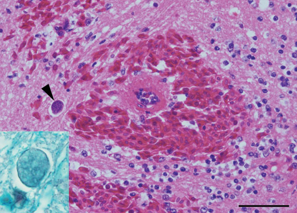
Figure. Acute severe encephalomalacia in the caudal brainstem of a captive Yellow-spotted bell frog from Sydney, Australia. This lesion was characterized by hemorrhage, vascular necrosis, and parasites consistent with Myxozoa (arrowhead) (hematoxylin and eosin stain; scale bar = 50 μm). Staining for axons confirmed intraaxonal location of the myxozoan parasites (inset, Holmes silver nitrate with Loxul Fast Blue stain).
Page created: March 16, 2012
Page updated: March 16, 2012
Page reviewed: March 16, 2012
The conclusions, findings, and opinions expressed by authors contributing to this journal do not necessarily reflect the official position of the U.S. Department of Health and Human Services, the Public Health Service, the Centers for Disease Control and Prevention, or the authors' affiliated institutions. Use of trade names is for identification only and does not imply endorsement by any of the groups named above.