Atypical Scrapie Prions from Sheep and Lack of Disease in Transgenic Mice Overexpressing Human Prion Protein
Jonathan D.F. Wadsworth

, Susan Joiner, Jacqueline M. Linehan, Anne Balkema-Buschmann, John Spiropoulos, Marion M. Simmons, Peter C. Griffiths, Martin H. Groschup, James Hope, Sebastian Brandner, Emmanuel A. Asante, and John Collinge
Author affiliations: University College London, London, UK (J.D.F Wadsworth, S. Joiner, J.M. Linehan, S. Brandner, E.A. Asante, J. Collinge); Federal Research Institute for Animal Health, Greifswald-Insel Riems, Germany (A. Anne Balkema-Buschmann, M.H. Groschup); Animal Health and Veterinary Laboratories Agency, Addlestone, UK (J. Spiropoulos, M.M. Simmons, P.C. Griffiths, J. Hope)
Main Article
Figure 3
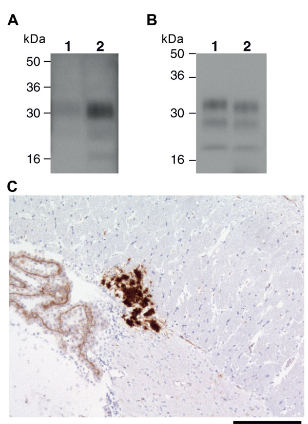
Figure 3. . Ovine bovine spongiform encephalopathy (BSE) prion transmission to a 129MM Tg35c mouse. Panel A shows immunoblot detection of disease-related prion protein (PrPSc) in 10 μL of proteinase K (PK)–digested 10% (w/v) brain homogenates from ovine BSE (SE 1929/0877) (lane 1) and secondary passage ovine BSE (SE1945/0032) (lane 2) using monoclonal antibody ICSM35 against prion protein (PrP). Panel B shows type 4 PrPSc in 1 μL of PK-digested 10% (w/v) vCJD brain homogenate (lane 1) in comparison to PrPSc in 20 μL of PK-digested 10% (w/v) brain homogenate from a 129MMTg35c mouse with subclinical prion infection that was culled 661 days after inoculation with secondary passage ovine BSE inoculum SE1945/0032 (lane 2). Panel C shows abnormal PrP immunoreactivity stained with monoclonal antibody ICSM35 against PrP in the corpus callosum of the ovine BSE–affected 129MM Tg35c mouse brain. Scale bar indicates 165 μm.
Main Article
Page created: October 31, 2013
Page updated: October 31, 2013
Page reviewed: October 31, 2013
The conclusions, findings, and opinions expressed by authors contributing to this journal do not necessarily reflect the official position of the U.S. Department of Health and Human Services, the Public Health Service, the Centers for Disease Control and Prevention, or the authors' affiliated institutions. Use of trade names is for identification only and does not imply endorsement by any of the groups named above.