Volume 20, Number 4—April 2014
Dispatch
Burkholderia pseudomallei Type G in Western Hemisphere
Jay E. Gee , Christopher J. Allender, Apichai Tuanyok, Mindy G. Elrod, and Alex R. Hoffmaster
Figure 1
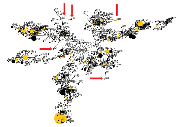
Figure 1. . Diagram of eBURST (3) analysis of multilocus sequence types (STs) of Burkholderia pseudomallei isolates; default settings were used. Dots represent sequence types. Blue dot represents the calculated primary founder, yellow dots represent calculated subgroup founders, and black dots represent the remaining STs. The sizes of dots are proportional to the number of isolates in the database representing a given ST. A single line between sequence types indicates that they are single-locus variants of each other. Red arrows indicate positions of STs associated with isolates from the Western Hemisphere and that are type G according to internal transcribed spacer typing.
References
- Wiersinga WJ, Currie BJ, Peacock SJ. Melioidosis. N Engl J Med. 2012;367:1035–44 . DOIPubMedGoogle Scholar
- Godoy D, Randle G, Simpson AJ, Aanensen DM, Pitt TL, Kinoshita R, Multilocus sequence typing and evolutionary relationships among the causative agents of melioidosis and glanders, Burkholderia pseudomallei and Burkholderia mallei. J Clin Microbiol. 2003;41:2068–79. DOIPubMedGoogle Scholar
- Feil EJ, Li BC, Aanensen DM, Hanage WP, Spratt BG. eBURST: inferring patterns of evolutionary descent among clusters of related bacterial genotypes from multilocus sequence typing data. J Bacteriol. 2004;186:1518–30. DOIPubMedGoogle Scholar
- Pearson T, Giffard P, Beckstrom-Sternberg S, Auerbach R, Hornstra H, Tuanyok A, Phylogeographic reconstruction of a bacterial species with high levels of lateral gene transfer. BMC Biol. 2009;7:78 . DOIPubMedGoogle Scholar
- Liguori AP, Warrington SD, Ginther JL, Pearson T, Bowers J, Glass MB, Diversity of 16S–23S rDNA internal transcribed spacer (ITS) reveals phylogenetic relationships in Burkholderia pseudomallei and its near-neighbors. PLoS ONE. 2011;6:e29323. DOIPubMedGoogle Scholar
- Tuanyok A, Auerbach RK, Brettin TS, Bruce DC, Munk AC, Detter JC, A horizontal gene transfer event defines two distinct groups within Burkholderia pseudomallei that have dissimilar geographic distributions. J Bacteriol. 2007;189:9044–9. DOIPubMedGoogle Scholar
Page created: March 18, 2014
Page updated: March 18, 2014
Page reviewed: March 18, 2014
The conclusions, findings, and opinions expressed by authors contributing to this journal do not necessarily reflect the official position of the U.S. Department of Health and Human Services, the Public Health Service, the Centers for Disease Control and Prevention, or the authors' affiliated institutions. Use of trade names is for identification only and does not imply endorsement by any of the groups named above.