Volume 21, Number 3—March 2015
Letter
Rickettsial Infections in Monkeys, Malaysia
To the Editor: The cynomolgus monkey (Macaca fascicularis), also known as the long-tailed macaque or crab-eating monkey, is commonly found in the Southeast Asia region (1). The macaque has been associated with several bacterial infections, such as those caused by hemotropic Mycoplasma and Bartonella quintana (2). As a result of rapid deforestation and changes in land use patterns, cynomolgus monkeys live in close proximity to human-populated areas (1). Human–macaque conflict may increase the risk for zoonoses.
Little is known about rickettsial and anaplasma infections in cynomolgus monkeys in Malaysia. Although Rickettsia spp. RF2125 and Rf31 have been identified from cat fleas in Malaysia (3), the presence of Anaplasma bovis in monkeys is not known.
Rickettsia felis, a member of the spotted fever group rickettsiae, is an emergent fleaborne human pathogen distributed worldwide (4). The obligate intracellular bacterium has been identified from cats, dogs, opossums, and the ectoparasites of various mammalian hosts. Several uncultured rickettsiae genetically closely related to the R. felis–type strain URRWXCal2 (referred to as R. felis–like organisms and including Rickettsia spp. RF2125, Rf31, Candidatus Rickettsia asemboensis, and others) have also been identified from various arthropods and fecal samples of primates (5). A. bovis is a gram-negative, pleomorphic, tickborne intracellular bacterium that infects a wide range of mammal species in many geographic regions (6).
To learn more about these infections in monkeys, we examined blood samples from 50 cynomolgus monkeys caught by the Department of Wildlife and National Parks at 12 residential areas in Peninsular Malaysia during a population management and wildlife disease surveillance program (January 2012–December 2013). Most monkeys (14 male, 36 female) were adults and were active and healthy. DNA was extracted from 200 μL of each blood sample by using a QIAamp DNA Mini Kit (QIAGEN, Hilden, Germany). We performed PCRs selective for the rickettsial citrate synthase gene (gltA) by using primers CS-78 and CS-323 and for the 135-kDa outer membrane protein B gene (ompB) by using primers 120-M59 and 120-807 (7). As positive controls, we used cloned PCR4-TOPO TA plasmids (Invitrogen, Carlsbad, CA, USA) with amplified gltA fragment from R. honei (strain TT118) and ompB fragment from a rickettsial endosymbiont (98% similarity to R. raoultii) of a tick sample. Amplification of anaplasma DNA was performed by using a group-specific primer pair (EHR 16SD/EHR 16SR) (8). As a positive control for the PCR, we used an A. marginale–infected cattle blood sample. The full-length sequences of the Anaplasma 16S rRNA gene were obtained by amplification with primers ATT062F and ATT062R (9). Sequence determination of the amplicons was performed by using forward and reverse primers of respective PCRs on an ABI PRISM 377 Genetic Analyzer (Applied Biosystems, Waltham, MA, USA). To search for homologous sequences in the GenBank database, we performed a BLAST (http://blast.ncbi.nlm.nih.gov/Blast.cgi) analysis and constructed a dendrogram based on 16S rDNA sequences of A. bovis (10).
The rickettsial gltA gene was detected from 12 (24%) blood samples of mostly male monkeys from 8 locations. BLAST analysis of 210 nucleotides (GenBank accession no. KP126803) amplified from all samples demonstrated 100% sequence similarity with Rickettsia sp. RF2125 (accession no. AF516333), Candidatus Rickettsia asemboensis (accession no. JN315968), and Rickettsia spp. clone 4G/JP102 and 11TP21 (accession nos. JN982949 and JN982950), which had been identified from cat fleas in Southeast Asia, Africa, and Costa Rica, respectively. The rickettsial sequence also showed 99.0% similarity (2-nt difference) with R. felis–type strain (accession no. CP000053). The rickettsial ompB gene was amplified from 4 samples, and BLAST analysis of the sequences (556–779 bp) revealed closest match to several R. felis–like organisms, including Rickettsia sp. RF2125 (100%, accession no. JX183538) and Candidatus Rickettsia asemboensis (99%, accession no. JN315972). BLAST analysis of the longest ompB sequence (accession no. KP126804) obtained in this study showed 93% similarity with that of the R. felis–type strain.
Figure
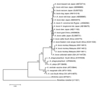
Figure. Phylogenetic relationships among various Anaplasma species, based on partial sequences of the 16S rRNA gene (1,263 bp). The dendrogram was constructed by using the neighbor-joining method in MEGA6 software (
Anaplasma DNA was amplified from 5 (10%) monkeys at 2 locations by using group-specific primers. Analysis of the nearly full-length sequences of the A. bovis 16S rRNA gene (1,457 nt) revealed 3 sequence types (GenBank accession nos. KM114611–3) with 99.1%–99.2% homology to that of the A. bovis strain from cattle in South Africa (accession no. U03775). The phylogenetic tree (Figure) inferred by using various Anaplasma species confirms the clustering of the strains from monkeys with A. bovis from different animals (i.e., goats, cattle, deer, ticks, wild boars, dogs, raccoons, leopard cats, eastern rock sengis, and cottontail rabbits). Co-infection of R. felis–like organisms and A. bovis was detected in only 1 sample.
Infections caused by R. felis–like organisms and A. bovis in the cynomolgus monkeys were subclinical (i.e., monkeys showed no evident signs of infection at the time of blood sampling). The diverse range of the organisms’ ectoparasite and animal hosts raises concern about their potential risk to human and animal health. Further study on the interactions between the microbes, vectors, and reservoir hosts is needed to assess their effects on public health.
Acknowledgments
We thank the Department of Wildlife and National Parks Peninsular Malaysia, Kuala Lumpur, Malaysia, for permission to conduct this study (reference no. JPHL&TN[IP]-80-4/2Jilid15[51]).
This project was funded by grants HIR-MOHE E000013-20001 (subprogramme 4), UM.C/625/1/HIR-MOHE/CHAN/11, and UMRG RP013-2012A from the University of Malaya, Kuala Lumpur.
References
- Gumert MD. The common monkey of Southeast Asia: long-tailed macaque populations, ethnophoresy, and their occurrence in human environments. In: Fuentes A, Gumert M, Jones-Engel L, editors. Monkeys on the edge: ecology and management of long-tailed macaques and their interface with humans. Cambridge (UK): Cambridge University Press; 2011. p. 3–12.
- Maggi RG, Mascarelli PE, Balakrishnan N, Rohde CM, Kelly CM, Ramaiah L, “Candidatus Mycoplasma haemomacaque” and Bartonella quintana bacteremia in cynomolgus monkeys. J Clin Microbiol. 2013;51:1408–11. DOIPubMedGoogle Scholar
- Tay ST, Mokhtar AS, Low KC, Mohd Zain SN, Jeffery J, Abdul Aziz N, Identification of rickettsiae from wild rats and cat fleas in Malaysia. Med Vet Entomol. 2014;28(Suppl 1):104–8. DOIPubMedGoogle Scholar
- Parola P. Rickettsia felis: from a rare disease in the USA to a common cause of fever in sub-Saharan Africa. Clin Microbiol Infect. 2011;17:996–1000. DOIPubMedGoogle Scholar
- Odhiambo AM, Maina AN, Taylor ML, Jiang J, Richards AL. Development and validation of a quantitative real-time polymerase chain reaction assay specific for the detection of Rickettsia felis and not Rickettsia felis–like organisms. Vector Borne Zoonotic Dis. 2014;14:476–81 . DOIPubMedGoogle Scholar
- Dumler JS, Barbet AF, Bekker CP, Dasch GA, Palmer GH, Ray SC, Reorganization of genera in the families Rickettsiaceae and Anaplasmataceae in the order Rickettsiales: unification of some species of Ehrlichia with Anaplasma, Cowdria with Ehrlichia and Ehrlichia with Neorickettsia, descriptions of six new species combinations and designation of Ehrlichia equi and ‘HGE agent’ as subjective synonyms of Ehrlichia phagocytophila. Int J Syst Evol Microbiol. 2001;51:2145–65. DOIPubMedGoogle Scholar
- Krawczak FS, Nieri-Bastos FA, Nunes FP, Soares JF, Moraes-Filho J, Labruna MB. Rickettsial infection in Amblyomma cajennense ticks and capybaras (Hydrochoerus hydrochaeris) in a Brazilian spotted fever–endemic area. Parasit Vectors. 2014;7:7.
- Parola F, Roux V, Camicas JL, Baradji I, Brouqui P, Raoult D. Detection of ehrlichiae in African ticks by polymerase chain reaction. Trans R Soc Trop Med Hyg. 2000;94:707–8. DOIPubMedGoogle Scholar
- Pinyoowong D, Jittapalapong S, Suksawat F, Stich RW, Thamchaipenet A. Molecular characterization of Thai Ehrlichia canis and Anaplasma platys strains detected in dogs. Infect Genet Evol. 2008;8:433–8. DOIPubMedGoogle Scholar
- Tamura K, Stecher G, Peterson D, Filipski A, Kumar S. MEGA6: Molecular Evolutionary Genetics Analysis version 6.0. Mol Biol Evol. 2013;30:2725–9. DOIPubMedGoogle Scholar
Figure
Cite This ArticleRelated Links
Table of Contents – Volume 21, Number 3—March 2015
| EID Search Options |
|---|
|
|
|
|
|
|
Please use the form below to submit correspondence to the authors or contact them at the following address:
Sun Tee Tay, Department of Medical Microbiology, Faculty of Medicine, University of Malaya, Kuala Lumpur 50603, Malaysia
Top