Volume 5, Number 5—October 1999
Research
Abscesses due to Mycobacterium abscessus Linked to Injection of Unapproved Alternative Medication
Abstract
An unlicensed injectable medicine sold as adrenal cortex extract (ACE*) and distributed in the alternative medicine community led to the largest outbreak of Mycobacterium abscessus infections reported in the United States. Records from the implicated distributor from January 1, 1995, to August 18, 1996, were used to identify purchasers; purchasers and public health alerts were used to identify patients. Purchasers and patients were interviewed, and available medical records were reviewed. Vials of ACE* were tested for mycobacterial contamination, and the product was recalled by the U.S. Food and Drug Administration. ACE* had been distributed to 148 purchasers in 30 states; 87 persons with postinjection abscesses attributable to the product were identified. Patient and vial cultures contained M. abscessus identical by enzymatic and molecular typing methods. Unusual infectious agents and alternative health practices should be considered in the diagnosis of infections that do not respond to routine treatment.
Almost half of the U.S. population uses some form of unconventional therapy, most without the knowledge of their physician (1). Although many alternative therapies are not directly associated with adverse outcomes, unlicensed injectable preparations may pose a significant health risk. Outbreaks due to alternative therapies are particularly challenging to detect, investigate, and control. The difficulty is compounded when the adverse outcome is an unusual infection with a prolonged incubation period.
We report on a multistate outbreak of postinjection abscesses associated with the use of an injectable product purported to contain adrenal cortex extract (ACE). The product was widely distributed in the alternative medicine community. ACE has been in use since 1895, when William Osler reported success with a glycerol extract of fresh adrenal tissue in the treatment of Addison disease (2). In the 1930s, ACE became commercially available for the diagnosis and treatment of Addison disease and other states of adrenal insufficiency. Synthetic formulations of adrenal hormones replaced ACE in general use. Although ACE never received U.S. Food and Drug Administration (FDA) approval, it remains in use by alternative medicine practitioners for such conditions as alcohol and drug withdrawal, allergies, inflammation, and stress management, as well as hypoglycemia and depression attributed to a state of "hypoadrenalism" (3,4).
The Mycobacterium abscessus outbreak was detected after two Denver-area physicians reported soft-tissue abscesses in patients who received injections of a product purported to contain ACE. We determined that the cause of the outbreaks was intrinsic contamination with M. abscessus of vials of the product from one distributor (this product is hereafter referred to as ACE*); ACE* was recalled by FDA. This article describes the investigation of the distribution and purchasers of ACE*; the conditions treated with ACE*; and the occurrences, of abscesses as well as the treatment and clinical course of affected patients.
Epidemiologic Investigation
Passive surveillance was enhanced, and patients and practitioners were alerted of health risks associated with ACE* (5). We requested invoices for purchases of ACE* from the implicated Arizona-based distributor for January 1, 1995, to August 18, 1996. Purchasers were contacted by telephone, informed of ACE*'s possible contamination, and asked to submit remaining unopened and opened vials for culture. A case-patient was defined as a person who had received an injection of ACE* between January 1, 1995, and August 18, 1996, and had developed an abscess at the site of injection.
Purchaser and Patient Questionnaires
A standardized questionnaire was used to collect information about the purchasers of ACE*, their use of the product, and any adverse outcomes. Purchasers who prescribed the product to others or were involved in further distribution were asked to identify persons who received ACE*.
Persons who received injections of ACE* were identified through the distribution list, health-care providers, and pharmacies that dispensed ACE* directly to patients. Other patients contacted health departments after learning of the outbreak through news reports. We completed a standardized questionnaire by reviewing the medical record (whenever possible) or by interviewing the patient on the telephone. When a practitioner declined to disclose the patient's identity, we interviewed the practitioner to complete the patient questionnaire. Data collected included demographic information; conditions treated with ACE*; dosage, route, and frequency of ACE* administration; location of ACE* injection, in a practitioner's office or at home; complications; dates of injections, abscess onset, and seeking of medical care; treatments given; and dates of abscess resolution. If dosage, frequency, or dates were not documented, the best estimate of the provider or patient was recorded. When only a range of doses, frequencies, or dates was given, the midpoint was selected.
Laboratory Investigation
All purchasers and patients contacted by CDC were asked to submit opened and unopened vials of ACE*. FDA also collected vials for analysis and submitted acid-fast isolates cultured from vials to CDC for identification and subtyping. Clinical laboratories also sampled vials for contamination and sent isolates from vials as well as patients to the National Jewish Medical and Research Center. Isolates identified as M. abscessus were forwarded to CDC for subtyping.
Mycobacterial cultures were performed by aspirating 5 ml of fluid from each vial of ACE*; 0.1 ml was plated onto Middlebrook and Cohn 7H10 agar with OADC enrichment (Difco Laboratories, Detroit, MI), and the remainder was used to inject 40 ml of Middlebrook 7H9 broth with ADC enrichment (Difco). Mycobacterial isolates were grown and identified by methods previously described (6,7).
Isolates were typed by multilocus enzyme electrophoresis (MEE) and pulsed-field gel electrophoresis (PFGE) (8,9). All isolates of mycobacteria and control strains were typed by MEE. PFGE was used to compare three randomly selected outbreak isolates of M. abscessus (two vial isolates, one patient isolate) and 27 random control isolates for their large restriction fragment patterns with the endonucleases Xba I and Dra I (9,10).
Selected isolates of M. abscessus were tested for susceptibility to amikacin, kanamycin, and tobramycin with disk diffusion; cefoxitin and minocycline with agar elution; and imipenem, ciprofloxacin, and clarithromycin with broth microdilution (11). Three outbreak isolates of M. abscessus and controls were tested for susceptibility to mercury (12).
The Manufacturing Process and the Product
An investigation of the manufacturing process of the product was undertaken by FDA (13). The product was analyzed at the FDA Forensic Chemistry Center in Cincinnati, Ohio.
Abscesses Following ACE* Administration
Figure 2
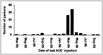
Figure 2. Dates of last injection of a presumed adrenal cortex extract among persons who developed postinjection Mycobacterium abscessus abscesses, United States, January 1995 to September 1996.
Figure 3
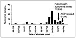
Figure 3. Dates of abscess onset in persons who had postinjection Mycobacterium abscessus abscesses after using a presumed adrenal cortex extract, United States, January 1995 to September 1996.
We identified 140 persons (treated in 20 states) who received ACE* during the interval of interest. Persons who received ACE* injections were 15 to 77 years of age (median 43 years, n = 131); 123 (88%) were women; 87 (62%) persons from 16 states had abscesses (Figure 1); 3% had other complications: fever and chills after injection (n = 2), acute reaction requiring intravenous fluids (n = 1), and a tender, swollen nodule at the site of a previous ACE* injection that did not meet our case definition (n = 1); 42 persons reported using ACE* without adverse effects (median follow-up 117 days, range 49-770 days); and data for 7 persons were lacking or insufficient. The Table compares case-patients with persons who reported that they did not develop an abcess after using ACE*. The dates of last injection of ACE* and onset of abscesses are shown in Figures 2 and 3, respectively. Most patients with abscesses had received intramuscular injections, though one had received an intravenous injection; most (n = 77, 89%) had received injections in a health-provider's office. Of the 35 case-patients who received only a single injection of ACE*, time to noticeable development of an abscess or documentation of the abscess in the medical record, whichever was earlier, was 4 to 149 days (median 32 days).
Treatment and Natural History of Abscesses
Sixty (69%) case-patients received medical care for the abscesses, and some received more than one type of therapy: 51 (59%) had incision and drainage (14 more than once), 41 (47%) were prescribed an antibiotic, and 11 (13%) required surgical excision or plastic surgery. Abscesses were cultured for mycobacteria in 21 (24%) case-patients, 12 of whom were patients of the same practitioner. Treatment was delayed in many cases. Thirty-four (39%) case-patients received either an incision and drainage procedure or a prescription for an antibiotic active against M. abscessus within 6 weeks of developing a noticeable abscess. Treatment courses of clarithromycin, in this outbreak the most commonly prescribed antibiotic having activity against M. abscessus (14), lasted a median of 30 days, (range 4-210 days, n = 19). Data were not sufficient to allow a comparison of treatment regimens.
Time to abscess resolution was estimated by using the earliest reported or documented date of the complication and the date on which the abscess had resolved (on the basis of the medical record or the patient's interpretation of resolution). Follow-up data were available on 42 (48%) case-patients. Eleven (13%) patients reported complete resolution of abscesses during the study period. Abscesses lasted 31 to 428 days (median 167 days) in the 10 persons for whom we have dates of onset and resolution. Abscesses persisted in 31 (36%) case-patients (median follow-up interval 217 days, range 22-672 days, n = 30). The outcome of the remaining 45 case-patients could not be ascertained.
Administration of ACE* by Health-Care Providers
Of 103 health-care providers who purchased ACE*, 58 (56%) were medical doctors, 19 (18%) doctors of osteopathy, 17 (17%) alternative practitioners (9 naturopaths, 6 chiropractors, 1 practitioner of homeopathic medicine, 1 holistic practitioner); the qualifications of 9 (9%) providers could not be determined. Providers used ACE* most commonly for chronic fatigue syndrome (n = 39), "hypoadrenalism" (n = 34), immune system enhancement, or infection (n = 11). One physician used ACE* extensively for weight loss. Dose per injection was 0.1 to 15 cc (median 2 cc), most commonly as a weekly injection (n = 31 [36%]) but ranged from a single injection to daily injections. Providers (n = 80) estimated treating a median of 7 patients each with ACE* (1 to several hundred) in the preceding 2 years. In some cases the provider's reported practice differed markedly from the quantity of ACE* ordered. One practitioner reported treating 12 patients with ACE* with an average of 3.5 ml per dose on a weekly to monthly basis, but invoices recorded that 410 (30 ml) vials of ACE* were shipped to this practitioner over the 20-month period.
Most providers [n = 91] administered ACE* in their offices (n = 77 [85%]), although others provided ACE* to their patients for home administration. Providers (n = 89) injected ACE* intramuscularly (n = 38 [43%]), intravenously (n = 35 [39%]), subcutaneously (n = 2 [2%]), or by more than one route (n = 14 [16%]). When given intravenously, ACE* was commonly (n = 42 [47%]) mixed with other injectable preparations including vitamins, minerals, and (in a few cases) crude liver extract.
Laboratory Results
From at least 38 purchasers, CDC and FDA obtained 248 vials labeled ACE. Of these, 213 vials were tested for mycobacterial contamination (177 unopened and 36 opened vials). M. abscessus was cultured from 7 vials (6 unopened), M. mucogenicum from 17 unopened vials, and both from 1 unopened vial. The 11 patient isolates of M. abscessus were identical to the 8 vial isolates of M. abscessus by MEE but differed from control isolates. Three outbreak isolates identical by MEE (data not shown) and PFGE (Figure 4) differed from 27 control isolates. These 3 isolates were resistant to mercury (as were the isolates of M. mucogenicum) but susceptible to clarithromycin, imipenem, amikacin, and kanamycin; moderately susceptible to cefoxitin; and resistant to ciprofloxacin, tobramycin, and minocycline.
Tracing ACE* Distribution
All implicated vials were from the same Arizona-based distributor. This distributor provided 337 invoices, representing shipment of 3,954 vials (each containing 30 ml of ACE*) to 148 purchasers between January 1, 1995, and August 18, 1996. ACE* was shipped to 30 states and two foreign countries. Of 146 U.S. purchasers, 103 (71%) were health-care providers, 8 (6%) were pharmacies or pharmaceutical companies, 11 (8%) were persons who purchased ACE* for self-administration; the remaining 24 (16%) could not be reached or declined to be interviewed. Purchasers received shipments of 1 to 200 vials (median 6). During the period of interest, one Dallas-based company received 13 shipments totaling 700 30-ml vials; the company declined to provide information about further distribution. We were able to trace 2,702 (68%) vials distributed to either health-care providers or persons who self-administered ACE*.
Manufacturing Process and Product Components
According to court documents, ACE* was manufactured in Florida under nonsterile conditions using a handwritten formula. The formula specified a low concentration of hydrocortisone mixed with the powdered preservative merthiolate (an organomercurial) dissolved in distilled water. Samples of the original distilled water were not available for testing. The product was placed in unlabeled vials and shipped to Montana, where labels bearing the names of nonexistent pharmaceutical companies were added. Vials contained 163 µg/ml to 234 µg/ml hydrocortisone and were found to contain mercury. There is no evidence that adrenal glands or other animal products were used to produce ACE* (13).
Wide distribution of an unlicensed injectable preparation contaminated with M. abscessus led to a multistate outbreak of soft-tissue abscesses. The outbreak went undetected for longer than 1 year before cases were reported to public health authorities. Reporting led to a nationwide investigation and recall of the implicated product.
Nonsterile water has been suspected or implicated in most reported outbreaks of M. abscessus. Outbreaks have followed cardiac surgery (15), cosmetic surgery (16), podiatric procedures (17), invasive otologic procedures (18), and dialysis (19,20). In most outbreaks of postinjection abscesses due to rapidly growing nontuberculous mycobacteria, multidose vials appear to have become contaminated and served as a common source (21-23). Multidose vials of lidocaine were suspected in a recent large outbreak in which 350 patients given injections by an alternative medicine practitioner in Colombia developed abscesses or skin lesions (24,25). In this outbreak, distilled water used to dissolve the preservative merthiolate may have been the source of contamination. Although approximately 80% of M. abscessus isolates are susceptible to mercury (12), patient and vial isolates from this outbreak were uniformly resistant, which would explain their ability to grow in the presence of merthiolate. M. mucogenicum was cultured from unopened vials of ACE* but not from patient abscesses; however, in most cases abscesses were not cultured.
Despite a growing number of reports, M. abscessus and other mycobacteria remain an underappreciated cause of postinjection abscesses, particularly those of long incubation that are unresponsive to antibiotics active against more common bacterial species. If special culturing techniques and prolonged incubation of culture media are not performed, the diagnosis may not be established, and appropriate treatment may be delayed or omitted. In this outbreak, mycobacteria were infrequently considered by the treating physician as a possible cause of the abscess. Only one fourth of case-patients were documented to have had mycobacterial cultures of an abscess; half of these patients were under the care of a single practitioner. Less than one third of patients with a postinjection abscess received an antibiotic active against mycobacteria, and most of those received an abbreviated course unlikely to have effected cure.
Randomized treatment trials of cutaneous disease, including localized abscesses, due to M. abscessus are lacking. Pretreatment isolates are susceptible to the oral antimicrobial agent clarithromycin (14), and most are susceptible to the parenterally administered antibiotics cefoxitin, imipenem, amikacin, and kanamycin (11); however, variations in resistance patterns make it imperative to determine the drug susceptibilities of each clinical isolate. In the largest reported outbreak of postinjection abscesses, the combination of surgical excision with clarithromycin appeared more efficacious (53% resolution at 3 months) than either treatment method alone (32% and 23%, respectively) (25). Optimal therapy would include two oral antimicrobial agents combined with incision and drainage. When only one oral antimicrobial agent is active against M. abscessus, combining incision and drainage with clarithromycin monotherapy may be practicable in cases of localized soft-tissue infection. This strategy was not associated with the development of complications in this outbreak. Although monotherapy with clarithromycin has been shown to select for resistance in disseminated M. chelonae (26-29) and disseminated M. abscessus infections (29), the risk appears lower in localized disease. Clarithromycin should be continued for at least 3 to 6 months or longer depending on clinical response (25,28).
Tracing of contaminated vials of ACE* was hampered not only by the wide initial distribution and the secondary distribution by some recipients but also by the lack of lot numbers and expiration dates on the labels. We discovered 87 case-patients through distribution invoices and public alerts but this number probably underestimates the extent of the outbreak. The role of ACE* in the development of abscesses was underappreciated or overlooked by patients and their health-care providers: eight patients continued to receive injections of ACE* after abscesses developed; one of these received injections 8 months after the abscess formed.
Recent interest in assessing the efficacy of alternative medical practices in a scientifically rigorous manner (30,31) serves the public's health by differentiating between potentially beneficial medicines and ineffectual or dangerous compounds. This report underscores not only the dangers posed to public health by unlicensed products from manufacturers not following good manufacturing practices but also the delays that can result in investigating an outbreak due to such products. Unusual infectious agents and alternative health practices should be considered in the diagnosis of infections that do not respond to routine treatment.
Dr. Galil is a medical epidemiologist with the National Immunization Program, CDC.
Acknowledgment
We thank the many patients and health-care providers who provided information, William R. Mac Kenzie for his thoughtful review of the manuscript, Pam Lindholm-Levy for antimicrobial susceptibility testing, Barbara Brown for performing broth microdilution and mercury susceptibility testing, and Yansheng Zhang for performing PFGE. The investigation would not have been possible without the assistance of Lynn Weaver, the cooperation of FDA, and many state and local health departments. We also thank David Ashford, Ulana Bodnar, Kristy Bradley, Mary Anne Caine, Lisa Cairns, Debra Chew, Sara Cody, Todd Damrow, David Dassey, Annie Fine, Jesse Greenblatt, Richard Hoffman, Daniel Jernigan, Daniel Jorgensen, J.P. Lofgren, Laurene Mascola, Gayle Miller, Craig Nichols, Pekka Nuorti, Barbara Ray, Larry Shireley, Kathleen Toomey, Andre Weltman, and Kenneth Wilcox.
References
- Eisenberg DM, Davis RB, Ettner SL, Appel S, Wilkey S, Van Rompay M, Trends in alternative medicine use in the United States. JAMA. 1988;280:1569–75. DOIGoogle Scholar
- Osler W. On six cases of Addison's Disease with the report of a case greatly benefited by the use of suprarenal extract. International Medical Magazine. 1895;4:3–11.
- Eggleston CW. Soma. Adrenal cortical hormone therapy. Am J Med Sci. 1939;197:718–29. DOIGoogle Scholar
- Hypoglycemia Association. 1998. Bulletin #96. Available from: URL: http://www.fred.net/slowup/hai96.html.
- Infection with. Mycobacterium abscessus associated with intramuscular injection of adrenal cortex extract—Colorado and Wyoming, 1995-1996. MMWR Morb Mortal Wkly Rep. 1996;45:713–5.PubMedGoogle Scholar
- Smithwick RW, Bigbie MR Jr, Ferguson RB, Karlix MA, Wallis CK. Phenolic acridine orange fluorescent stain for mycobacteria. J Clin Microbiol. 1995;33:2763–4.PubMedGoogle Scholar
- Butler WR, Jost KC Jr, Kilburn JO. Identification of mycobacteria by high-performance liquid chromatography. J Clin Microbiol. 1991;29:2468–72.PubMedGoogle Scholar
- Yakrus MA, Reeves MW, Hunter SB. Characterization of isolates of Mycobacterium avium serotypes 4 and 8 from patients with AIDS by multilocus enzyme electrophoresis. J Clin Microbiol. 1992;30:1474–8.PubMedGoogle Scholar
- Wallace RJ Jr, Zhang Y, Brown BA, Fraser V, Mazurek GH, Maloney S. DNA large restriction fragment patterns of sporadic and epidemic nosocomial strains of Mycobacterium chelonae and Mycobacterium abscessus. J Clin Microbiol. 1993;31:2697–701.PubMedGoogle Scholar
- Tenover FC, Arbeit RD, Goering RV, Mickelsen PA, Murray BE, Persing DH, Interpreting chromosomal DNA restriction patterns produced by pulsed-field gel electrophoresis: criteria for bacterial strain typing. J Clin Microbiol. 1995;33:2233–9.PubMedGoogle Scholar
- Swenson JM, Wallace RJ Jr, Silcox VA, Thornsberry C. Antimicrobial susceptibility of five subgroups of Mycobacterium fortuitum and Mycobacterium chelonae [published erratum appears in Antimicrob Agents Chemother 1986 Apr;29:720]. Antimicrob Agents Chemother. 1985;28:807–11.PubMedGoogle Scholar
- Steingrube VA, Wallace RJ Jr, Steele LC, Pang YJ. Mercuric reductase activity and evidence of broad-spectrum mercury resistance among clinical isolates of rapidly growing mycobacteria. Antimicrob Agents Chemother. 1991;35:819–23.PubMedGoogle Scholar
- U.S. District Court. Southern District of Florida. Complaint for preliminary and permanent injunction. Miami (FL): The Court; 1997. CIV-Case 97-1083.
- Brown BA, Wallace RJ Jr, Onyi GO, De Rosas V, Wallace RJ III. Activities of four macrolides, including clarithromycin, against Mycobacterium fortuitum, Mycobacterium chelonae, and M. chelonae-like organisms. Antimicrob Agents Chemother. 1992;36:180–4.PubMedGoogle Scholar
- Kuritsky JN, Bullen MG, Broome CV, Silcox VA, Good RC, Wallace RJ Jr. Sternal wound infections and endocarditis due to organisms of the Mycobacterium fortuitum complex. Ann Intern Med. 1983;98:938–9.PubMedGoogle Scholar
- Safranek TJ, Jarvis WR, Carson LA, Cusick LB, Bland LA, Swenson JM, Mycobacterium chelonae wound infections after plastic surgery employing contaminated gentian violet skin-marking solution. N Engl J Med. 1987;317:197–201.PubMedGoogle Scholar
- Wenger JD, Spika JS, Smithwick RW, Pryor V, Dodson DW, Carden GA, Outbreak of Mycobacterium chelonae infection associated with use of jet injectors. JAMA. 1990;264:373–6. DOIPubMedGoogle Scholar
- Lowry PW, Jarvis WR, Oberle AD, Bland LA, Silberman R, Bocchini JA Jr, Mycobacterium chelonae causing otitis media in an ear-nose-and-throat practice. N Engl J Med. 1988;319:978–82.PubMedGoogle Scholar
- Bolan G, Reingold AL, Carson LA, Silcox VA, Woodley CL, Hayes PS, Infections with Mycobacterium chelonei in patients receiving dialysis and using processed hemodialyzers. J Infect Dis. 1985;152:1013–9.PubMedGoogle Scholar
- Lowry PW, Beck-Sague CM, Bland LA, Aguero SM, Arduino MJ, Minuth AN, Mycobacterium chelonae infection among patients receiving high-flux dialysis in a hemodialysis clinic in California. J Infect Dis. 1990;161:85–90.PubMedGoogle Scholar
- Inman PM, Beck A, Brown AE, Stanford JL. Outbreak of injection abscesses due to Mycobacterium abscessus. Arch Dermatol. 1969;100:141–7. DOIPubMedGoogle Scholar
- Borghans JG, Stanford JL. Mycobacterium chelonei in abscesses after injection of diphtheria-pertussis-tetanus-polio vaccine. Am Rev Respir Dis. 1973;107:1–8.PubMedGoogle Scholar
- Gremillion DH, Mursch SB, Lerner CJ. Injection site abscesses caused by Mycobacterium chelonei. Infect Control. 1983;4:25–8.PubMedGoogle Scholar
- Camargo D, Saad C, Ruiz F, Ramirez ME, Lineros M, Rodriguez G, Iatrogenic outbreak of M. chelonae skin abscesses. Epidemiol Infect. 1996;117:113–9. DOIPubMedGoogle Scholar
- Villanueva A, Calderon RV, Vargas BA, Zhang Y, Sander P, Onyi GO, Report on an outbreak of postinjection abscesses due to Mycobacterium abscessus, including management with surgery and clarithromycin therapy and comparison of strains by random amplified polymorphic DNA polymerase chain reaction. Clin Infect Dis. 1997;24:1147–53. DOIPubMedGoogle Scholar
- Tebas P, Sultan F, Wallace RJ Jr, Fraser V. Rapid development of resistance to clarithromycin following monotherapy for disseminated Mycobacterium chelonae infection in a heart transplant patient. Clin Infect Dis. 1995;20:443–4.PubMedGoogle Scholar
- Driscoll MS, Tyring SK. Development of resistance to clarithromycin after treatment of cutaneous Mycobacterium chelonae infection. J Am Acad Dermatol. 1997;36:495–6. DOIPubMedGoogle Scholar
- Wallace RJ Jr, Tanner D, Brennan PJ, Brown BA. Clinical trial of clarithromycin for cutaneous (disseminated) infection due to Mycobacterium chelonae. Ann Intern Med. 1993;119:482–6.PubMedGoogle Scholar
- Wallace RJ Jr, Meier A, Brown BA, Zhang Y, Sander P, Onyi GO, Genetic basis for clarithromycin resistance among isolates of Mycobacterium chelonae and Mycobacterium abscessus. Antimicrob Agents Chemother. 1996;40:1676–81.PubMedGoogle Scholar
- Alternative medicine: a report to the National Institutes of Health on alternative medical systems and practices in the United States, Workshop on Alternative Medicine; 1992 Sep 14-16; Chantilly, Virginia. Washington: U.S. Government Printing Office; 1993.
- Villaire M. OAM report: centers provide OAM with `deliverables'. J Altern Ther. 1997;3:20–4. DOIGoogle Scholar
Figures
Table
Cite This ArticleTable of Contents – Volume 5, Number 5—October 1999
| EID Search Options |
|---|
|
|
|
|
|
|
Please use the form below to submit correspondence to the authors or contact them at the following address:
Karin Galil, Centers for Disease Control and Prevention, 1600 Clifton Road, Mail Stop E61, Atlanta, Georgia 30333, USA; fax: 404-639-8616
Top