Volume 6, Number 4—August 2000
Dispatch
Evidence of Infection in Humans with Rickettsia helvetica in Eastern France
Figure 2
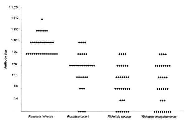
Figure 2. Antibody titers against Rickettsia helvetica, R. conorii, R. slovaca, and "R. mongolotimonae" in 35 forest workers, Alsace.* * Each diamond represents the IgG titer in serum samples from the patients. R. conorii Moroccan strain (ATCC VR 141), R. helvetica C9P9 strain, R. slovaca 13-B strain, and "R. mongolotimonae" HA91 strain were grown on confluent layers of Vero cells in a 150-cm2 flask. Infection was monitored by Gimenez staining. When 90% of the cells were infected, cell layers and supernatants were harvested, pelleted by centrifugation (10,000 x g for 10 min), and resuspended in 15 mL of phosphate-buffered saline (PBS, pH 7.3). Antigen purification was performed (5). The final suspension was resuspended in sterile water, and the protein content of the purified organisms was determined by UV spectrophotometry and adjusted to 1 mg/mL. All four rickettsial antigens were applied by pen point to each well of 30-well microscope slides (Dynatech Laboratories Ltd., Billingshurt, United Kingdom), air dried, and fixed with acetone for 10 min. This allowed a direct comparison of fluorescence intensity between antigens. All serum samples, including positive and negative controls, were diluted 1/4, 1/8, 1/16, 1/32, 1/64 and 1/128 in PBS with 3% nonfat dry milk. Microimmunofluorescence procedures were performed (3). All serum samples found to be positive for total immunoglobulins were diluted serially (twofold dilutions ranging from 1/32 to 1/2,048 or more), and the titers of IgG and IgM were determined. Before detection of IgM, serum was adsorbed with a rheumatoid factor absorbent (RF-absorbent, Behring-werke AG, Marburg, Germany). Titers at or above 1:64 for IgG or 1:32 for IgM were considered positive.
References
- Gilot B. Bases biologiques, écologiques et cartographiques pour l'étude des maladies transmises par les tiques (Ixodidae et Argasidae) dans les alpes françaises et leur avant pays) [thesis]. Grenoble, France: Universite de Grenoble; 1985.
- Nilsson K, Lindquist O, Pahlson C. Association of Rickettsia helvetica with chronic perimyocarditis in sudden cardiac death. Lancet. 1999;354:1169–73. DOIPubMedGoogle Scholar
- Brouqui P, Harle JR, Delmont J, Frances C, Weiller PJ, Raoult D. African tick bite fever : an imported spotless rickettsiosis. Arch Intern Med. 1997;157:119–24. DOIPubMedGoogle Scholar
- Teysseire N, Raoult D. Comparison of Western immunoblotting and microimmunofluoresence for diagnosis of Mediterranean spotted fever. J Clin Microbiol. 1992;30:455–60.PubMedGoogle Scholar
- Raoult D, Dasch GA. Line blot and Western blot immunoassays for diagnosis of Mediterranean spotted fever. J Clin Microbiol. 1989;27:2073–9.PubMedGoogle Scholar
- Hechemy KE, Raoult D, Fox J, Han Y, Elliott LB, Rawlings J. Cross-reaction of immune sera from patients with rickettsial diseases. J Med Microbiol. 1989;29:199–202. DOIPubMedGoogle Scholar