Accumulation of L-type Bovine Prions in Peripheral Nerve Tissues
Yoshifumi Iwamaru

, Morikazu Imamura, Yuichi Matsuura, Kentaro Masujin, Yoshihisa Shimizu, Yujing Shu, Megumi Kurachi, Kazuo Kasai, Yuichi Murayama, Shigeo Fukuda, Sadao Onoe, Ken’ichi Hagiwara, Yoshio Yamakawa, Tetsutaro Sata, Shirou Mohri, Hiroyuki Okada, and Takashi Yokoyama
Author affiliations: National Institute of Animal Health, Tsukuba, Ibaraki, Japan (Y. Iwamaru, M. Imamura, Y. Matsuura, K. Masujin, Y. Shimizu, Y. Shu, M. Kurachi, K. Kasai, Y. Murayama, S. Mohri, H. Okada, T. Yokoyama); Hokkaido Animal Research Center, Hokkaido, Japan (S. Fukuda, S. Onoe); National Institute of Infectious Diseases, Tokyo, Japan (K. Hagiwara, Y. Yamakawa, T. Sata)
Main Article
Figure 1
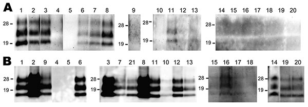
Figure 1. Western blot analysis of a protease-resistant form (PrPres) of a normal cellular prion protein in nerve tissue samples obtained from cattle 10 (A) and 16 (B) months postinoculation (cattle identification codes 8515 and 1061, respectively). The nerve tissues tested are shown above the lanes: 1, trigeminal ganglia; 2, pituitary gland; 3, anterior cervical ganglion; 4, facial nerve; 5, hypoglossal nerve; 6, cranial mesenteric ganglia; 7, vagus nerve (cervical part); 8, stellate ganglia; 9, adrenal gland; 10, phrenic nerve; 11, vagus nerve (pectoral part); 12, vagosympathic trunk (pectoral part); 13, vagosympathetic trunk (lumbar part); 14, accessory nerve; 15, suprascapular nerve; 16, brachial nerve plexus; 17, median nerve; 18, radial nerve; 19, sciatic nerve; 20, tibial nerve, 21, middle cervical ganglion. The equivalent of 100 mg of tissue was loaded. Western blots were probed with monoclonal antibody T2 to detect PrPres. Molecular mass standards (kDa) are indicated on the left of each panel.
Main Article
Page created: March 02, 2011
Page updated: March 02, 2011
Page reviewed: March 02, 2011
The conclusions, findings, and opinions expressed by authors contributing to this journal do not necessarily reflect the official position of the U.S. Department of Health and Human Services, the Public Health Service, the Centers for Disease Control and Prevention, or the authors' affiliated institutions. Use of trade names is for identification only and does not imply endorsement by any of the groups named above.