Skip directly to site content Skip directly to page options Skip directly to A-Z link Skip directly to A-Z link Skip directly to A-Z link
Volume 17, Number 3—March 2011
Research

Amplification of Emerging Viruses in a Bat Colony

Jan Felix Drexler1, Victor Max Corman1, Tom Wegner, Adriana Fumie Tateno, Rodrigo Melim Zerbinati, Florian Gloza-Rausch, Antje Seebens, Marcel A. Müller, and Christian DrostenComments to Author 
Author affiliations: University of Bonn Medical Centre, Bonn, Germany (J.F. Drexler, V.M. Corman, A.F. Tateno, R.M. Zerbinati, F. Gloza-Rausch, A. Seebens, M.A. Müller, C. Drosten); University of Bonn, Bonn (T. Wegner); Noctalis, Centre for Bat Protection and Information, Bad Segeberg, Germany (F. Gloza-Rausch, A. Seebens)

Main Article

Figure 3

Detection frequency of bat viruses and virus nucleic acid concentrations over time. A) Coronavirus; B) astrovirus; C) adenovirus. Samples were obtained approximately every 3 weeks from the same Myotis myotis bat maternity roost in 3 different sampling years, 2008–2010. Each sample was tested by specific real-time reverse transcription–PCR (RT-PCR) with RNA/DNA concentrations per gram of feces given on the y axis. The arrows indicate the time of birth of the first pup. Numbers on the x-axis repre

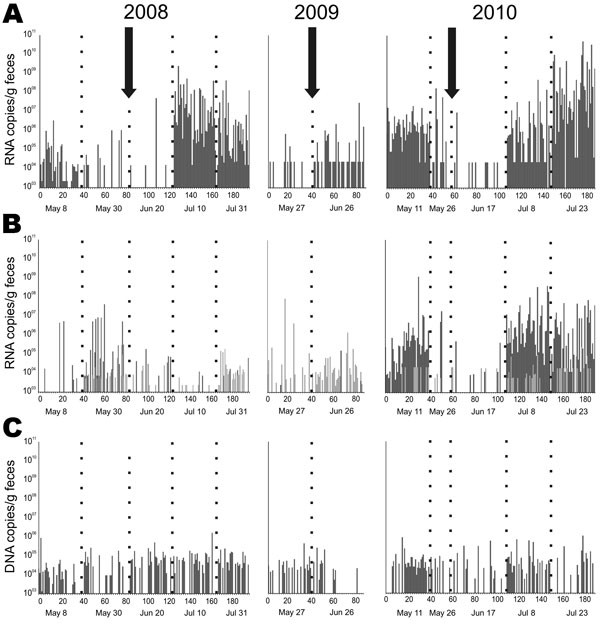
Figure 3. Detection frequency of bat viruses and virus nucleic acid concentrations over time. A) Coronavirus; B) astrovirus; C) adenovirus. Samples were obtained approximately every 3 weeks from the same Myotis myotis bat maternity roost in 3 different sampling years, 2008–2010. Each sample was tested by specific real-time reverse transcription–PCR (RT-PCR) with RNA/DNA concentrations per gram of feces given on the y axis. The arrows indicate the time of birth of the first pup. Numbers on the x-axis represent individual fecal pools tested, consisting of 5 single fecal pellets each. Five different sampling dates (below each panel) are shown by dotted lines for each sampling year. Empty columns indicate pools that tested negative. In panel B, light and dark gray bars identify results by 2 different real-time RT-PCRs that were used simultaneously to cover the large astrovirus diversity encountered.

Main Article

1These authors contributed equally to this article.

Page created: January 24, 2018
Page updated: January 24, 2018
Page reviewed: January 24, 2018
The conclusions, findings, and opinions expressed by authors contributing to this journal do not necessarily reflect the official position of the U.S. Department of Health and Human Services, the Public Health Service, the Centers for Disease Control and Prevention, or the authors' affiliated institutions. Use of trade names is for identification only and does not imply endorsement by any of the groups named above.
file_external