Volume 10, Number 3—March 2004
Research
Coronaviridae and SARS-associated Coronavirus Strain HSR1
Elisa Vicenzi* , Filippo Canducci*, Debora Pinna*, Nicasio Mancini*, Silvia Carletti*, Adriano Lazzarin*†, Claudio Bordignon*†, Guido Poli*†, and Massimo Clementi*†
Figure 3
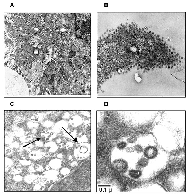
Figure 3. Ultrastructural analysis of Vero cells infected with severe acute respiratory syndrome–associated coronavirus (SARS-CoV) strain HSR1. A, intracellular budding of SARS-CoV in large vesicles containing CoV virions (magnification x30,000); B, clusters of extracellular virions adjacent to the plasma membrane (magnification x50,000); C and D, intracellular budding of SARS-CoV virions (magnification x50,000).
Page created: February 08, 2011
Page updated: February 08, 2011
Page reviewed: February 08, 2011
The conclusions, findings, and opinions expressed by authors contributing to this journal do not necessarily reflect the official position of the U.S. Department of Health and Human Services, the Public Health Service, the Centers for Disease Control and Prevention, or the authors' affiliated institutions. Use of trade names is for identification only and does not imply endorsement by any of the groups named above.