Volume 13, Number 5—May 2007
Letter
Vancomycin-resistant Enterococci, Mexico City
Cite This Article
Citation for Media
To the Editor: Vancomycin-resistant Enterococcus (VRE) has become an important nosocomial pathogen because of its rapid spread, limited therapy options, mortality, and the possibility of transfer of vancomycin resistance to other pathogens such as Staphylococcus aureus. Vancomycin-resistant E. faecium (VREF) and E. faecalis were first described in 1988 (1,2).They have become major nosocomial pathogens, but their prevalence in Latin America has remained <2% (3). In Mexico, VRE has rarely been reported (4,5). In a recent study in Mexico City, 100% (n = 60) of the isolates of E. faecium and E. faecalis were susceptible to vancomycin (6).
From May 2004 to April 2005, the rate of vancomycin resistance among all Enterococcus isolates was 0.27%. However, in May 2005 the first fully VREF was isolated at our hospital, and the rate of vancomycin resistance was 6.23% (a 23-fold increase) during the following 12-month period.
We performed a retrospective study to describe the isolates and the characteristics of patients with VREF. All VREF isolates from May 2005 through April 2006 were included. We collected demographic and clinical data. For the final identification of the isolates, the VITEK system (bioMérieux, Lyon, France) with VITEK GPI cards (bioMérieux, Inc., Durham NC, USA) were used. Antimicrobial drug susceptibility was tested by using the VITEK GPS-111 card and confirmed by MIC determination that used broth microdilution. Resistance to vancomycin and teicoplanin was confirmed by E-test (AB Biodisk, Solna, Sweden). An isolate was considered vancomycin resistant when the MIC was ≥32 μg/mL and was considered to have high-level resistance when the MIC was ≥256 μg/mL. A PCR for detection of the vanA or vanB genotype was used (7). Isolates were characterized by pulsed-field gel electrophoresis (PFGE) (8,9); a dendrogram was constructed with the GelCompare II 4.0 software (Applied Maths, Kortrijk, Belgium), and the similarity was compared with the Dice coefficient.
In the study period, VREF was isolated from 27 patients. The median age was 40 years (range 22–84 years). VREF was isolated from the abdomen in 14 patients (51.9%); 11 isolates were from an abscess, 2 from infected surgical sites, and 1 from ascites. An additional 8 isolates were from the urinary tract (29.6%), 2 from the bloodstream (7.4%), 2 from soft-tissue (7.4%), and 1 (3.7%) from bone. Residence in the general medical wards during the isolation of VREF was most common, 17 (63%) cases, followed by 6 (22.2%) in the intensive care unit. The remaining 4 (14.8%) were distributed in other areas. Median time of hospitalization before the isolation was 21 days (range 1–84 days). Twenty-five patients (92.6%) had a central line, 12 (44.4%) had mechanical ventilation, and 20 (74.1%) previous surgery. Of the last group, 17 (85%) of 20 had abdominal surgery. Twenty-four patients (88.8%) received an antimicrobial drug before the isolation of VREF: third- or fourth-generation cephalosporins (89%), metronidazole (70.4%), aminoglycosides (70.4%), vancomycin (66.7%), carbapenems (66.7%), amoxicillin or ampicillin (48.1%), antifungal agents (48.1%); and <20% received quinolones, trimethoprim-sulfamethoxazole, colistin, macrolides, and antimycobacterial or antiviral agents. The median time of antimicrobial drug use was 11 days (range 1–84 days). During hospitalization, 7 patients died (crude death rate, 25.9%), 5 of them from sepsis with at least another microorganism isolated; the remaining 2 died of gastrointestinal hemorrhage.
All isolates of E. faecium had a vancomycin MIC ≥256 μg/mL and a vanA phenotype (teicoplanin resistance); 26 (96.3%) had vanA genotype. Only 1 isolate of E. faecium was classified as non–vanA, non vanB, even though it demonstrated high-level resistance to vancomycin and teicoplanin. Resistance to other antimicrobial agents was as follows: ampicillin and ciprofloxacin, 100%; high-level gentamicin, 48.2%; quinupristin/dalfopristin, 7.4%; and linezolid, 0%.
Figure
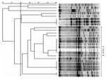
Figure. Pulsed-field gel electrophoresis (PFGE) banding patterns of chromosomal DNA of 26 isolates of vancomycin-resistant enterococci. There is a clear predominant type, classified as type A (≥80% similarity), composed of 18 isolates...
PFGE analysis showed several genotypes of E. faecium; however, 18 of 26 of the isolates had <3 band differences from the predominant strain classified as type A. One isolate of E. faecium could not be typed (Figure).
As in most tertiary-care centers, our PFGE data suggest that a heterogenous population of VREF exists, but a particular clone established itself as the dominant strain. Although infection control measures are well established in our hospital, in disseminated outbreaks caused by several different clones, infection control measures and control of vancomycin use have shown only limited efficacy. This suggests selection pressure by antimicrobial drugs other than vancomycin (10). Early detection of VREF is of extreme importance because of the possibility that the vanA gene may be transferred to a variety of gram-positive microorganisms, including S. aureus.
The rate of isolation of VREF at our hospital increased considerably during the last year. Even though the number of patients is small, we consider this finding to be of utmost importance, since VREF seems to be emerging in Mexico. To our knowledge, this is the first well-documented outbreak of high-level resistance to vancomycin in enterococci in Mexico. Further research is needed to determine if the problem is limited to our hospital or if it is a nationwide trend.
References
- Uttley AH, Collins CH, Naidoo J, George RC. Vancomycin-resistant enterococci [letter]. Lancet. 1988;l:57–8. DOIGoogle Scholar
- Centers for Disease Control and Prevention. Nosocomial enterococci resistant to vancomycin—United States, 1989–1993. MMWR Morb Mortal Wkly Rep. 1993;42:597–9.PubMedGoogle Scholar
- Low DE, Keller N, Barth A, Jones RN. Clinical prevalence, antimicrobial susceptibility, and geographic resistance patterns of enterococci: results from the SENTRY Antimicrobial Surveillance Program, 1997–1999. Clin Infect Dis. 2001;32(Suppl 2):S133–45. DOIPubMedGoogle Scholar
- Sifuentes-Osornio J, Ponce-de-León A, Muñoz-Trejo T, Villalobos-Zapata Y, Ontiveros-Rodriguez C, Gómez-Roldan C. Antimicrobial susceptibility patterns and high-level gentamicin resistance among enterococci isolated in a Mexican tertiary care center. Rev Invest Clin. 1996;48:91–6.PubMedGoogle Scholar
- McDonald LC, Garza LR, Jarvis WR. Proficiency of clinical laboratories in and near Monterrey, Mexico, to detect vancomycin-resistant enterococci. Emerg Infect Dis. 1999;5:143–6.PubMedGoogle Scholar
- Cornejo-Juarez P, Velásquez-Acosta C, Díaz-Gonzalez A, Volkow-Fernandez P. Tendencia del perfil de sensibilidad antimicrobiana de los aislamientos de sangre en un hospital oncológico (1998–2003). Salud Publica Mex. 2005;47:288–93. DOIPubMedGoogle Scholar
- Dutka-Malen S, Evers S, Courvalin P. Detection of glycopeptide resistance genotypes and identification to the species level of clinically relevant enterococci by PCR. J Clin Microbiol. 1995;33:24–7.PubMedGoogle Scholar
- Miranda AG, Singh KV, Murray BE. DNA fingerprinting of Enterococcus faecium by pulse-field gel electrophoresis may be a useful epidemiologic tool. J Clin Microbiol. 1991;29:2752–7.PubMedGoogle Scholar
- Tenover FC, Arbeit RD, Goering RV, Mickelsen PA, Murray BE, Persing DH, Interpreting chromosomal DNA restriction patterns produced by pulsed-field gel electrophoresis: criteria for bacterial strain typing. J Clin Microbiol. 1995;33:2233–9.PubMedGoogle Scholar
- Rice LB. Emergence of vancomycin-resistant enterococci. Emerg Infect Dis. 2001;7:183–7.PubMedGoogle Scholar
Figure
Cite This ArticleRelated Links
Table of Contents – Volume 13, Number 5—May 2007
| EID Search Options |
|---|
|
|
|
|
|
|
Please use the form below to submit correspondence to the authors or contact them at the following address:
José Sifuentes-Osornio, Instituto Nacional de Ciencias Médicas y Nutrición, Salvador Zubirán, Vasco de Quiroga No. 15, Col. Sección XVI, Del. Tlapan, México, DF CP 14000, México;
Top