Volume 13, Number 6—June 2007
Dispatch
Full Recovery from Baylisascaris procyonis Eosinophilic Meningitis
Poulomi J. Pai*, Brian G. Blackburn†1, Kevin R. Kazacos‡, Rajasekharan P. Warrier*, and Rodolfo E. Bégué*
Figure
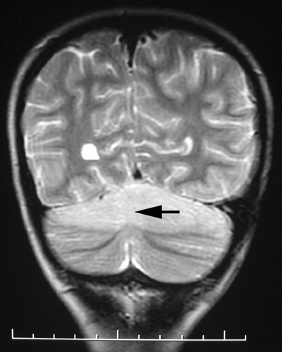
Figure. Coronal T2-weighted magnetic resonance imaging of the brain in a 4-year-old child with Baylisascaris procyonis eosinophilic meningitis. Arrow shows diffuse edema of the superior cerebellar hemispheres. Scale bar increments = cm.
Page created: May 31, 2011
Page updated: May 31, 2011
Page reviewed: May 31, 2011
The conclusions, findings, and opinions expressed by authors contributing to this journal do not necessarily reflect the official position of the U.S. Department of Health and Human Services, the Public Health Service, the Centers for Disease Control and Prevention, or the authors' affiliated institutions. Use of trade names is for identification only and does not imply endorsement by any of the groups named above.