Volume 14, Number 1—January 2008
Dispatch
Avian Influenza Virus (H5N1) Replication in Feathers of Domestic Waterfowl
Yu Yamamoto* , Kikuyasu Nakamura*, Masatoshi Okamatsu*, Manabu Yamada*, and Masaji Mase*
Figure 2
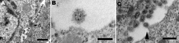
Figure 2. Pathology of a duck infected with A/chicken/Yamaguchi/7/2004. A) Electron microscopy of the feather epidermis showing virions observed between epidermal cells with the desmosome (d) and nucleus (n). Bar = 500 nm. B) Spherical virion with envelope spikes. Bar = 100 nm. C) Budding process of virion (arrowhead). Bar = 250 nm.
Page created: July 08, 2010
Page updated: July 08, 2010
Page reviewed: July 08, 2010
The conclusions, findings, and opinions expressed by authors contributing to this journal do not necessarily reflect the official position of the U.S. Department of Health and Human Services, the Public Health Service, the Centers for Disease Control and Prevention, or the authors' affiliated institutions. Use of trade names is for identification only and does not imply endorsement by any of the groups named above.