Volume 14, Number 4—April 2008
Research
Emericella quadrilineata as Cause of Invasive Aspergillosis
Cite This Article
Citation for Media
Abstract
We noted a cluster of 4 cases of infection or colonization by Emericella spp., identified by sequence-based analysis as E. quadrilineata. Sequence-based analysis of an international collection of 33 Emericella isolates identified 12 as E. nidulans, all 12 of which had previously been identified by morphologic methods as E. nidulans. For 12 isolates classified as E. quadrilineata, only 6 had been previously identified accordingly. E. nidulans was less susceptible than E. quadrilineata to amphotericin B (median MICs 2.5 and 0.5 mg/L, respectively, p<0.05); E. quadrilineata was less susceptible than E. nidulans to caspofungin (median MICs, 1.83 and 0.32 mg/L, respectively, p<0.05). These data indicate that sequence-based identification is more accurate than morphologic examination for identifying Emericella spp. and that correct species demarcation and in vitro susceptibility testing may affect patient management.
The genus Aspergillus includes >250 species; ≈20 have been reported to cause opportunistic infections in humans. The most important human pathogens in this genus are A. fumigatus, A. flavus, A. niger, A. terreus, and Emericella nidulans (anamorph: A. nidulans) (1). A. fumigatus is the most common cause of invasive aspergillosis, a condition associated with substantial severity and mortality rates (2). Invasive infections caused by E. nidulans are uncommon in animals and humans (3–5); in humans they appear to occur predominantly in patients who have chronic granulomatous disease (CGD), a rare disorder of phagocytes in which the absence of both superoxide and hydrogen peroxide production in phagocytes predisposes patients to bacterial and fungal infections. Invasive E. nidulans infections in this patient group are associated with higher mortality rates than those caused by A. fumigatus (6,7). The most common site of infection is the lungs; other manifestations are subcutaneous abscesses or liver abscesses, suppurative adenitis, osteomyelitis, fungemia, cellulitis, and meningitis (7,8). Within the genus Emericella, other species have only rarely been identified as agents of human or animal infections.
The identification of E. nidulans in clinical microbiology laboratories is commonly based on the characteristic microscopic morphology, the production of hülle cells, or the production of ascospores. A. fumigatus is identified by its heat tolerance; other species fail to grow when incubated at high temperature, typically 48°C.
We recently noted a cluster of infection or colonization by E. quadrilineata, a species closely related to E. nidulans. Within a 3-month period, 4 cases were identified at the Radboud University Nijmegen Medical Center. No apparent epidemiologic link between the cases was found because each patient was cared for in a different ward, and 2 patients with invasive aspergillosis were admitted directly from home. One of the latter 2 patients was a 10-year-old boy with X-linked CGD and a probable diagnosis of invasive pulmonary aspergillosis (9); the other patient was a 60-year-old man who had chronic lymphocytic leukemia and in whom cerebral aspergillosis later developed and was confirmed by biopsy. From the other 2 patients, who had no signs and symptoms of invasive fungal disease, E. quadrilineata was cultured from respiratory specimens. No laboratory contamination was evident during the period in which the cluster occurred. No subsequent cases were identified. Morphologic species identification was difficult, and we had to rely on sequence-based identification, which prompted this investigation of the role of E. quadrilineata as a causative agent of invasive aspergillosis.
Data and Strain Collection
We searched the PubMed literature for cases of infections caused by E. quadrilineata (anamorph: Aspergillus tetrazonus) or E. nidulans; search terms were Emericella, quadrilineata, tetrazonus, nidulans, and aspergillosis. For those articles that described infections by E. quadrilineata or E. nidulans, we asked the authors to send us their isolates for sequence analysis. We also approached colleagues who care for patients with CGD or might otherwise have a collection of E. nidulans isolates. We also searched our department’s fungal culture collection for E. nidulans isolates. It is our policy to store all Aspergillus isolates cultured from clinical specimens sent to our laboratory, regardless of the clinical relevance of the isolate. Finally, we added E. nidulans and E. quadrilineata isolates deposited in the culture collection of the Centraal Bureau voor Schimmelcultures ([CBS], Utrecht, the Netherlands). The final collection totaled 33 Emericella isolates, with 11 isolates from the CBS culture collection (type strains E. quadrilineata, E. nidulans, E. nidulans var. echinulata) and 1 isolate from the National Collection of Pathogenic Fungi. Ten isolates were from our own culture collection (including the 4 encountered in the cluster of cases), and 11 isolates were from 5 other medical centers; some of these isolates had been cultured as causes of infection and previously reported (online Appendix, available from www.cdc.gov/EID/content/14/4/566-appT.htm) (8,9–12). Seven isolates had been cultured from patients with confirmed invasive aspergillosis, and 2 were from patients with probable cases (online Appendix, available from www.cdc.gov/EID/content/14/4/566-appT.htm).
Morphologic Identification
Aspergillus isolates are routinely identified by their macroscopic colony morphology and the microscopic morphology of their anamorphic features. When teleomorph features were also used to identify an isolate, teleomorph nomenclature, such as Emericella spp., was used to report the strain. In addition, the isolates were incubated at 48ºC, which precludes the growth of most Aspergillus spp. except A. fumigatus.
Sequence-based Identification
Sequence-based identification in the routine clinical microbiology laboratory was carried out by sequencing of parts of the internal transcribed spacer (ITS) 1 and 2 regions. Total DNA of the Emericella cultures was extracted by using the MagNa Pure Total NA isolation kit (Roche Diagnostics Nederland BV, Almere, the Netherlands). Then the ITS 1 and 2 sequence was amplified by PCR with primers ITS1 (5′-TCCGTAGGTGAACCTGCGG-3′) and ITS4 (5′-TCCTCCGCTTATTGATATGC-3′) as described (13). After purification, the PCR product was sequenced with the BigDye Terminator v3 kit (Applied Biosystems, Foster City, CA, USA).
In addition to the above-mentioned sequenced-based identification, parts of the β-tubulin and calmodulin genes were sequenced. The Emericella cultures were cultivated in 2 mL malt peptone broth by using 10% (vol/vol) of malt extract (Oxoid, Basingstoke, UK) and 0.1% (wt/vol) bacto peptone (Difco, Becton Dickinson, Le Pont de Claix, France). The cultures were incubated at 25°C for 7 days. DNA was extracted from the cells by using the Masterpure yeast DNA purification kit (Epicentre Biotechnologies, Madison, WI, USA) according to the manufacturer’s instructions. Amplification of part of the β-tubulin gene was performed by using the primers Bt2a and Bt2b (14,15). Amplifications of the partial calmodulin gene were set up as described (16). Sequence analysis was performed with the BigDye Terminator Cycle Sequencing Ready Reaction Kit for both strands. Sequences were aligned by using ClustalX software (17) and were improved manually.
Evolutionary distances between the sequences were calculated by using the Kimura formula (18) and DNADIST program of the PHYLIP program package (19). Phylogenetic trees were prepared by using the neighbor-joining method (20) and the NEIGHBOR program of the PHYLIP package. Bootstrap values were calculated from 1,000 replications of the bootstrap procedure by using programs SEQBOOT, DNADIST, NEIGHBOR, and CONSENSE of the PHYLIP package (19,21). For parsimony analysis, PAUP* version 4.0 software was used (22). E. heterothallica was used as an outgroup in these experiments. The unique β-tubulin and calmodulin sequences were deposited in the GenBank nucleotide sequence database under accession nos. EF591677–EF591702.
Antifungal-Drug Susceptibility Testing
Antifungal-drug susceptibility testing of Emericella isolates was performed by using a microbroth dilution assay, as described by the Clinical Laboratory Standards Institute (M38-A) for amphotericin B (Bristol-Myers Squibb, Woerden, the Netherlands), itraconazole (Janssen-Cilag, Beerse, Belgium), voriconazole (Pfizer, Capelle aan den IJssel, the Netherlands), posaconazole (Schering-Plough, Maarsen, the Netherlands), caspofungin (MSD, Haarlem, the Netherlands), and terbinafine (Novartis, Arnhem, the Netherlands) (23). MICs were determined for all drugs except caspofungin, for which a microscopic endpoint was used (minimum effective concentration) (24). All in vitro susceptibility testing was performed in duplicate.
Statistical Analysis
After being transformed logarithmically, MIC dilutions were compared by using the Mann-Whitney U test. Data on growth at different antifungal drug concentrations were normalized by setting the corrected optical density of the growth control at 0% and the lowest optical density at 100%. Growth characteristics were analyzed by nonlinear regression analysis that used a 4-parameter logistic model and created a sigmoidal curve. Test runs determined deviation of the model, and goodness-of-fit was tested by determining r2 values. In addition to comparing MICs, we determined the drug concentration at which growth was 50% that of the control (50% maximal effective concentration [EC50]) and calculated and fitted the slope of the curve (GraphPad Prism, San Diego, CA, USA). For all drugs except caspofungin, the EC50 values and slopes were compared for E. nidulans and E. quadrilineata.
Species Identification
The 4 isolates from the cluster of cases grew on Sabouraud-dextrose agar as velvety, brownish-green colonies with a purplish reverse side. Conidiophores were light brown with hemispherical vesicles bearing metulae and biseriate philalides on the upper half. Conidia were spherical, smooth walled, subhyaline, finely roughened, and 3–4 μm in diameter. After ≈3 weeks of incubation, purple ascocarps formed, surrounded by characteristic hülle cells. Asci were spherical, 8 spored, 10–13 μm in diameter, and evanescent. The ascospores were reddish purple, lenticular, 5–6 × 3–4 μm, and smooth. The morphologic features were consistent with E. nidulans. As part of the diagnostic process, the isolates were incubated at 48ºC, and all isolates showed some growth, which was considered inconsistent with E. nidulans. Because of this discrepancy, sequence-based identification was performed. However, all 33 isolates from the subgenus Nidulantes section that were analyzed in this study grew at 48°C, which indicates that incubation at this temperature does not fully distinguish between this section and A. fumigatus.
Sequence-based Analysis
The ITS sequence analysis of the 4 isolates was consistent with that of E. quadrilineata, although there were only 1 or 2 mismatches with the base-pair sequence of E. nidulans. The morphologic features of E. nidulans and E. quadrilineata are very similar; only the microscopic shape of the ascospores shows subtle differences. Ascospores of E. nidulans have 2 longitudinal crests, as opposed to E. quadrilineata, which has 4 short equatorial crests. The resolution of the ITS region was considered too low to unambiguously differentiate between E. nidulans and E. quadrilineata, and further sequence-based identification was performed at CBS by using partial β-tubulin and calmodulin sequence data.
Figure 1
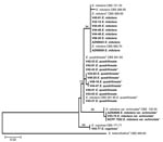
Figure 1. Neighbor-joining tree based on β-tubulin sequence data of the Emericella isolates examined. Clinical isolates are set in boldface. Numbers above branches are bootstrap values. Only values >70% are indicated. T indicates...
Figure 2
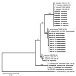
Figure 2. Neighbor-joining tree based on calmodulin sequence data of Emericella isolates examined. Clinical isolates are set in boldface. Numbers above branches are bootstrap values. Only values >70% are indicated. T indicates the...
During analysis of part of partial β-tubulin gene sequences, we analyzed 367 bases of all 33 isolates. Among the polymorphic sites, 23 were phylogenetically informative. The neighbor-joining tree (Figure 1) based on partial β-tubulin gene sequences had the same topologic features as 1 of the 2 maximum-parsimony trees constructed by the PAUP program (length 94 steps, consistency index 0.9787, retention index 0.9762). The calmodulin dataset included 489 bases, with 50 parsimony informative sites. The topologic features of the neighbor-joining tree (Figure 2) and 1 of the 2 most parsimonious trees were the same (tree length 162, consistency index 0.9691, retention index 0.9854). Molecular data indicated that 12 of 33 isolates could be classified as E. nidulans, all of which had previously been identified as E. nidulans by microscopic examination of morphologic characteristics or other methods. For the 12 isolates classified as E. quadrilineata, only 6 had previously been identified accordingly. These 6 isolates included the 4 in our cluster, 1 from the CBS culture collection, and 1 previously reported as the cause of onychomycosis (12). The remaining 6 isolates had been previously identified as E. nidulans (online Appendix, available from www.cdc.gov/EID/content/14/4/566-appT.htm). Of these, 1 belonged to the CBS culture collection, 1 was reported as the cause of cerebral aspergillosis (11), and 2 were from patients with CGD and confirmed invasive aspergillosis.
Figure 3
Figure 3. Scanning electron microscopic images of ascospores of some Emericella isolates. A) E. quadrilineata V43-63; B) E. rugulosa V43-77; C) E. nidulans var. echinulata 4606. Scale bars represent 5 μm.
A total of 4 isolates were classified as E. rugulosa, 1 of which had been previously reported as E. nidulans (10). A total of 4 isolates were identified as E. nidulans var. echinulata, 2 of which had caused invasive aspergillosis in patients with CGD and had been presumptively identified as E. nidulans. Scanning electron microscopy of the ascospores of some isolates supported their species assignment (JEOL 5600LV scanning electron microscope [JEOL, Tokyo, Japan] equipped with an Oxford CT1500 Cryostation [Oxford Instruments, Oxford, UK]) (Figure 3) (25).
In Vitro Susceptibility
The in vitro activity of antifungal agents against the Emericella isolates is shown in the online Appendix, available from www.cdc.gov/EID/content/14/4/566-appT.htm. Overall, terbinafine was the most active drug in vitro, followed by posaconazole, which was the most active azole. For statistical comparisons, we used 12 E. nidulans and 12 E. quadrilineata isolates. By comparing MICs, statistically significant differences in drug activity were found for amphotericin B, voriconazole, and posaconazole (Table). These differences were also found when EC50 values and slopes were compared for both species (data not shown). Comparison of minimum effective concentrations showed caspofungin to be significantly more active against the E. nidulans isolates (p<0.05). Although only 4 E. nidulans var. echinulata isolates were analyzed, the susceptibility profile of these isolates was similar to that of E. quadrilineata (and not of E. nidulans); for amphotericin B, MICs were low, and for caspofungin, MICs were high.
Literature Review
Three cases of infection due to E. quadrilineata have been documented. One patient was a 60-year-old man from northern India, who had a fingernail infection that affected all 5 nails of 1 hand. The strain was repeatedly cultured from 1 nail, and septate hyphal elements were seen in a portion of an excised nail. The patient was treated with itraconazole, but the response could not be evaluated (12). Invasive aspergillosis caused by E. quadrilineata has been described for 2 patients, both of whom had sinusitis. One of these, a 28-year-old woman, had acute nonlymphoblastic leukemia and had undergone allogeneic bone marrow transplantation. She developed sinusitis with orbital involvement 2 months after transplantation. The diagnosis was confirmed by biopsy, and the patient was successfully treated with a combination of surgical debridement, granulocyte transfusions, and intravenous amphotericin B–cholesterol sulfate colloidal dispersion (26). The other patient was a 28-year-old man who had received an allogeneic bone marrow transplant for acute myeloid leukemia. Left orbital swelling, facial pain, and nasal congestion developed 68 days after transplantation. E. quadrilineata was cultured from a biopsy specimen; the patient was successfully treated with external ethmoidectomy, granulocyte transfusions, and topical and systemic therapy with a lipid formulation of amphotericin B. The fungal infection resolved (27).
Until this report, 2 cases of invasive aspergillosis caused by E. quadrilineata had been described; each case had been reported as sinusitis in patients who had undergone bone marrow transplantation for hematologic malignancy. We add 1 case of central nervous system aspergillosis and 3 cases of invasive pulmonary aspergillosis in patients with CGD. The 3 cases may not be surprising because E. quadrilineata is very closely related to E. nidulans, a fungus known to cause infections in humans (3,5,28–30), primarily in patients with CGD (6,7). Emericella spp. other than E. nidulans are less frequently reported as causative agents of infectious disease. In addition to the above-mentioned human infections, E. quadrilineata has been identified as a causative agent of mycosis in animals (31). We have found only 1 report each of E. rugulosa (4) and E. nidulans var. echinulata (32) as the cause of human or animal infections.
Discriminating E. nidulans and E. quadrilineata by morphologic characteristics is virtually impossible. Only the ascospore ornamentation differs, and the subtle differences cannot be seen by using light microscopy. The most frequently used technique for their unambiguous identification is scanning electron microscopic examination of the lining of the ascospores (25). However, fruiting bodies are usually formed after a rather long incubation period (1–2 weeks). And although E. nidulans was found to form cleistothecia in the human body (33), clinical isolates often lose their ability to form sexual reproductive structures and ascospores (34,35). Given these difficulties, we anticipate that reliance on phenotypic characteristics alone would cause misidentification of E. nidulans, rather than correct identification of E. quadrilineata, as the cause of invasive aspergillosis. The sequence-based analysis showed that this was indeed the situation; 50% of the E. quadrilineata isolates had previously been identified as E. nidulans. Among these was a case of cerebral aspergillosis, the second case observed in our cluster of cases. Despite the close morphologic and genetic relatedness between E. nidulans and E. quadrilineata, the activity of antifungal agents differed significantly, which supports the conclusion that biological differences exist between these species. The triazoles were active in vitro; posaconazole showed the greatest activity, which is also observed for most Aspergillus spp. Although significant differences were found for activity of voriconazole and posaconazole, these differences appear to be not clinically relevant, given the small differences in MIC values (Table). However, for amphotericin B and caspofungin, the observed differences in activity may be clinically important. Amphotericin B was less active against E. nidulans than against E. quadrilineata. In vitro resistance of E. nidulans against amphotericin B has been recognized (36), although the testing method has been shown to substantially affect the activity found (37). However, lack of activity of amphotericin B has also been reported in experimental models of infection and in cases reported in the literature (8,38). Caspofungin was less active against E. quadrilineata than against E. nidulans. Caspofungin was shown to be effective against E. nidulans in a murine model of systemic infection (38), but no data are available for E. quadrilineata. Although the allylamine terbinafine is not used for treatment of patients with invasive aspergillosis, the drug is highly active against both E. nidulans and E. quadrilineata. The isolates were inhibited at concentrations as low as 0.015 mg/L.
Identification of molds primarily relies on morphologic criteria such as the macroscopic colony morphology and the microscopic morphology of the conidia and the structures bearing the conidia. Morphologic identification underestimates differences among species and among members of the same species. This was recently shown for the section Fumigati, in which A. lentulus and A. udagawae were among isolates phenotypically identified as A. fumigatus (34,35). We made a similar observation when 10 of 33 Emericella isolates were found to be misidentified. Correct species demarcation is important from a taxonomic viewpoint but can also have clinical relevance. Within the Aspergillus section of Fumigati, the newly identified species A. lentulus was shown to be more resistant than A. fumigatus to antifungal drugs (35). Therefore, correct species identification will affect the choice of antifungal therapy. Differences in drug activity were also apparent in the Emericella spp. we examined. Another important reason for correct species identification is the detection of outbreaks of infection, which could warrant interventions to prevent invasive fungal infection in immunocompromised persons or lead to epidemiologic surveys to identify sources of spread of fungal spores. However, the resolution of sequencing of the ITS region is too low to reliably differentiate between E. nidulans and E. quadrilineata; therefore, in vitro susceptibility testing might be appropriate in those laboratories that do not have access to sequencing of β-tubulin and calmodulin genes.
Molecular techniques in addition to morphologic identification have identified a role of E. quadrilineata as an opportunistic fungal pathogen, especially in patients with CGD and in those with hematologic malignancy. These molecular techniques will help identify and discriminate more accurately within the current fungal species and will give more insight into the pathogenesis of fungal infection.
Dr Verweij is professor of medical microbiology and chair of the Department of Medical Microbiology at Radboud University Nijmegen Medical Center. His special interest is invasive fungal diseases in immunocompromised patients. He has conducted research on the diagnosis of invasive aspergillosis, invasive candidiasis, and invasive zygomycosis, especially the performance of biological markers such as antigen and DNA.
Acknowledgment
We thank W. Steinbach, E. Roilides, E.M. Johnson, and J. Guarro for providing clinical isolates and relevant clinical information. We are grateful to R. Schade for helping collect relevant clinical and microbiologic information.
References
- Pagano L, Caira M, Picardi M, Candoni A, Melillo L, Fianchi L, Invasive aspergillosis in patients with acute leukemia: update on morbidity and mortality—SEIFEM-C report. Clin Infect Dis. 2007;44:1524–5. DOIPubMedGoogle Scholar
- Joshi KR, Mathur DR, Sharma JC, Vyas MC, Sanghvi A. Mycetoma caused by Aspergillus nidulans in India. J Trop Med Hyg. 1985;88:41–4.PubMedGoogle Scholar
- Knudtson WU, Kirkbride CA. Fungi associated with bovine abortion in the northern plains states (USA). J Vet Diagn Invest. 1992;4:181–5.PubMedGoogle Scholar
- Lucas GM, Tucker P, Merz WG. Primary cutaneous Aspergillus nidulans infection associated with a Hickman catheter in a patient with neutropenia. Clin Infect Dis. 1999;29:1594–6. DOIPubMedGoogle Scholar
- Dotis J, Roilides E. Osteomyelitis due to Aspergillus spp. in patients with chronic granulomatous disease: comparison of Aspergillus nidulans and Aspergillus fumigatus. Int J Infect Dis. 2004;8:103–10. DOIPubMedGoogle Scholar
- Winkelstein JA, Marino MC, Johnston RB Jr, Boyle J, Curnutte J, Gallin JI, Chronic granulomatous disease. Report on a national registry of 368 patients. Medicine. 2000;79:155–69. DOIPubMedGoogle Scholar
- Van ‘t Hek LG, Verweij PE, Weemaes CM, Van Dalen R, Yntema JB, Meis JF. Successful treatment with voriconazole of invasive aspergillosis in chronic granulomatous disease. Am J Respir Crit Care Med. 1998;157:1694–6.PubMedGoogle Scholar
- Berger PE, Warris A, Weemaes CM. Preventing fungal infections in chronic granulomatous disease. N Engl J Med. 2003;349:1190–1. DOIPubMedGoogle Scholar
- Dotis J, Panagopoulou P, Filioti J, Winn R, Toptsis C, Panteliadis C, Femoral osteomyelitis due to Aspergillus nidulans in a patient with chronic granulomatous disease. Infection. 2003;31:121–4. DOIPubMedGoogle Scholar
- Morris A, Schell WA, McDonagh D, Chaffee S, Perfect JR. Pneumonia due to Fonsecaea pedrosoi and cerebral abscesses due to Emericella nidulans in a bone marrow transplant recipient. Clin Infect Dis. 1995;21:1346–8.PubMedGoogle Scholar
- Gugnani HC, Vijayan KK, Tyagi P, Sharma S, Stchigel AM, Guarro J. Onychomycosis due to Emericella quadrilineata. J Clin Microbiol. 2004;42:914–6. DOIPubMedGoogle Scholar
- Henry T, Iwen PC, Hinrichs SH. Identification of Aspergillus species using internal transcribed spacer regions 1 and 2. J Clin Microbiol. 2000;38:1510–5.PubMedGoogle Scholar
- Glass NL, Donaldson GC. Development of primer sets designed for use with the PCR to amplify conserved genes from filamentous ascomycetes. Appl Environ Microbiol. 1995;61:1323–30.PubMedGoogle Scholar
- Samson RA, Houbraken JAMP, Kuijpers AFA, Frank JM, Frisvad JC. New ochratoxin or sclerotium producing species in Aspergillus section Nigri. Stud Mycol. 2004;50:45–61.
- Hong SB, Cho HS, Shin HD, Frisvad JC, Samson RA. Novel Neosartorya species isolated from soil in Korea. Int J Syst Evol Microbiol. 2006;56:477–86. DOIPubMedGoogle Scholar
- Thompson JD, Gibson TJ, Plewniak F, Jeanmougin F, Higgins DG. The ClustalX windows interface: flexible strategies for multiple sequence alignment aided by quality analysis tools. Nucleic Acids Res. 1997;25:4876–8. DOIPubMedGoogle Scholar
- Kimura M. A simple method for estimating evolutionary rates of base substitutions through comparative studies of nucleotide sequences. J Mol Evol. 1980;16:111–20. DOIPubMedGoogle Scholar
- Felsenstein J. PHYLIP (Phylogeny inference package), version 3.57c. Seattle: University of Washington; 1995.
- Saitou N, Nei M. The neighbor-joining method: a new method for reconstructing phylogenetic trees. Mol Biol Evol. 1987;4:406–25.PubMedGoogle Scholar
- Felsenstein J. Confidence limits on phylogenies: an approach using the bootstrap. Evolution Int J Org Evolution. 1985;39:783–91. DOIGoogle Scholar
- Swofford T. PAUP*: Phylogenetic analysis using parsimony, version 4.0. Sunderland (MA): Sinauer Associates; 2000.
- National Committee for Clinical Laboratory Standards. Reference method for broth dilution antifungal susceptibility testing of filamentous fungi. Approved standard M38-A. Wayne (PA): The Committee; 2002.
- Kurtz MB, Heath IB, Marrinan J, Dreikorn S, Onishi J, Douglas C. Morphological effects of lipopeptides against Aspergillus fumigatus correlate with activities against (1,3)-D-glucan synthase. Antimicrob Agents Chemother. 1994;38:1480–9.PubMedGoogle Scholar
- Horie Y. Ascospore ornamentation and its application to the taxonomic re-evaluation in Emericella [in Japanese]. Transactions of the Mycological Society of Japan. 1980;21:483–93.
- Polacheck I, Nagler A, Okon E, Drakos P, Plaskowitz J, Kwon-Chung KJ. Aspergillus quadrilineatus, a new causative agent of fungal sinusitis. J Clin Microbiol. 1992;30:3290–3.PubMedGoogle Scholar
- Drakos PE, Nagler A, Or R, Naparstek E, Kapelushnik J, Engelhard D, Invasive fungal sinusitis in patients undergoing bone marrow transplantation. Bone Marrow Transplant. 1993;12:203–8.PubMedGoogle Scholar
- Ng KP, Saw TL, Madasamy M, Soo Hoo T. Onychomycosis in Malaysia. Mycopathologia. 1999;147:29–32. DOIPubMedGoogle Scholar
- Tong QJ, Chai WX, Wang ZF, Kou JF, Qi ZT, Wang DL. A case of cerebral aspergillosis caused by Aspergillus nidulans. Clinical, pathologic and mycologic identifications. Chin Med J (Engl). 1990;103:518–22.PubMedGoogle Scholar
- Yano S, Kobayashi K, Shishido S, Nakano H. Intrabronchial Aspergillus nidulans infection in an immunocompetent man. Intern Med. 1999;38:372–5. DOIPubMedGoogle Scholar
- Singh MP, Singh CM. Fungi associated with suppurative mycosis of cattle and sheep in India. Indian J Anim Health. 1970;9:432–49.
- White CJ, Kwon-Chung KJ, Gallin JI. Chronic granulomatous disease of childhood. An unusual case of infection with Aspergillus nidulans var. echinulatus. Am J Clin Pathol. 1988;90:312–6.PubMedGoogle Scholar
- Mitchell RG, Chaplin AJ, Mackenzie DW. Emericella nidulans in a maxillary sinus fungal mass. J Med Vet Mycol. 1987;25:339–41. DOIPubMedGoogle Scholar
- Balajee SA, Gribskov J, Brandt M, Ito J, Fothergill A, Marr KA. Mistaken identity: Neosartorya pseudofischeri and its anamorph masquerading as Aspergillus fumigatus. J Clin Microbiol. 2005;43:5996–9. DOIPubMedGoogle Scholar
- Balajee SA, Nickle D, Varga J, Marr KA. Molecular studies reveal frequent misidentification of Aspergillus fumigatus by morphotyping. Eukaryot Cell. 2006;5:1705–12. DOIPubMedGoogle Scholar
- Kontoyiannis DP, Lewis RE, May GS, Osherov N, Rinaldi MG. Aspergillus nidulans is frequently resistant to amphotericin B. Mycoses. 2002;45:406–7. DOIPubMedGoogle Scholar
- Singh J, Rimek D, Kappe R. Intrinsic in vitro susceptibility of primary clinical isolates of Aspergillus fumigatus, Aspergillus terreus, Aspergillus nidulans, Candida albicans and Candida lusitaniae against amphotericin B. Mycoses. 2006;49:96–103. DOIPubMedGoogle Scholar
- Bowman JC, Abruzzo GK, Flattery AM, Gill CJ, Hickey EJ, Hsu MJ, Efficacy of caspofungin against Aspergillus flavus, Aspergillus terreus, and Aspergillus nidulans. Antimicrob Agents Chemother. 2006;50:4202–5. DOIPubMedGoogle Scholar
Figures
Table
Cite This ArticleTable of Contents – Volume 14, Number 4—April 2008
| EID Search Options |
|---|
|
|
|
|
|
|
Please use the form below to submit correspondence to the authors or contact them at the following address:
Paul E. Verweij, Radboud University Nijmegen Medical Center, Medical Microbiology, PO Box 9101, Nijmegen 6500 HB, the Netherlands;
Top