Volume 15, Number 5—May 2009
Research
Virulent Strain of Hepatitis E Virus Genotype 3, Japan
Kazuaki Takahashi , Hiroaki Okamoto, Natsumi Abe, Manri Kawakami, Hiroyuki Matsuda, Satoshi Mochida, Hiroshi Sakugawa, Yoshiki Suginoshita, Seishiro Watanabe, Kazuhide Yamamoto, Yuzo Miyakawa, and Shunji Mishiro
Appendix Figure
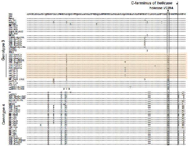
Appendix Figure. Alignment of a partial C-terminal amino acid sequence of the helicase domain of hepatitis E virus (HEV) open reading frame (ORF) 1 across genotype 3 and 4 isolates. A bracket indicates 8 isolates of the JIO strain HEV genotype 3 and 5 isolates of swJ19 strain. Conversions in helV239A are shown by linear box. Partial sequences of ORF1 (aa positions 1105-1226 of HEV-US2) of human and swine isolates of genotype 3 were compared.
Page created: December 16, 2010
Page updated: December 16, 2010
Page reviewed: December 16, 2010
The conclusions, findings, and opinions expressed by authors contributing to this journal do not necessarily reflect the official position of the U.S. Department of Health and Human Services, the Public Health Service, the Centers for Disease Control and Prevention, or the authors' affiliated institutions. Use of trade names is for identification only and does not imply endorsement by any of the groups named above.