Volume 15, Number 9—September 2009
Dispatch
Coxsackievirus A6 and Hand, Foot, and Mouth Disease, Finland
Riikka Österback, Tytti Vuorinen, Mervi Linna, Petri Susi, Timo Hyypiä, and Matti Waris
Figure 1
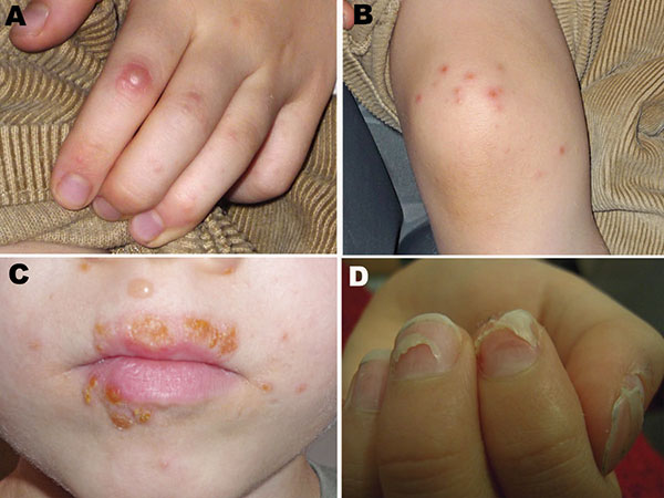
Figure 1. Vesicular eruptions in A) hand, B) foot, and C) mouth of a 6.5-year-old boy from Turku, Finland, with coxsackievirus (CV) A6 infection. Several of his fingernails shed 2 months after the pictures were taken. D) Onychomadesis in a 10-year-old boy from Seinäjoki, Finland, 2 months after hand, foot and mouth disease with CVA6 infection. Photographs courtesy of H. Kujari (A–C) and M. Linna (D).
Page created: December 07, 2010
Page updated: December 07, 2010
Page reviewed: December 07, 2010
The conclusions, findings, and opinions expressed by authors contributing to this journal do not necessarily reflect the official position of the U.S. Department of Health and Human Services, the Public Health Service, the Centers for Disease Control and Prevention, or the authors' affiliated institutions. Use of trade names is for identification only and does not imply endorsement by any of the groups named above.