Volume 16, Number 1—January 2010
Letter
Otomastoiditis Caused by Mycobacterium abscessus, the Netherlands
Jakko van Ingen , Frank Looijmans, Piet Mirck, Richard Dekhuijzen, Martin Boeree, and Dick van Soolingen
Appendix Figure
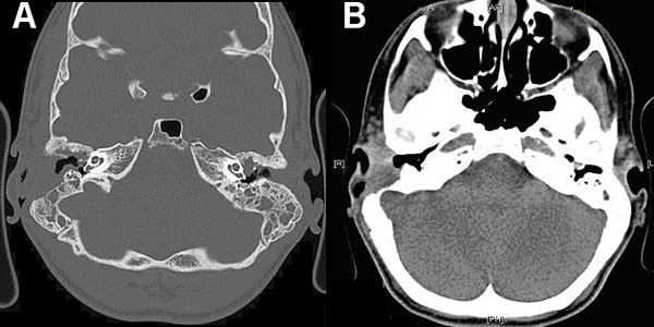
Appendix Figure. Computed tomography images of a patient with Mycobacterium abscessus otomastoiditis. Extensive bone destruction in the right mastoid and associated right-sided mucosal swelling can be seen. A) Bone tissue window setting; B) soft tissue window setting.
Page created: March 31, 2011
Page updated: March 31, 2011
Page reviewed: March 31, 2011
The conclusions, findings, and opinions expressed by authors contributing to this journal do not necessarily reflect the official position of the U.S. Department of Health and Human Services, the Public Health Service, the Centers for Disease Control and Prevention, or the authors' affiliated institutions. Use of trade names is for identification only and does not imply endorsement by any of the groups named above.