Skip directly to site content Skip directly to page options Skip directly to A-Z link Skip directly to A-Z link Skip directly to A-Z link
Volume 17, Number 3—March 2011
Dispatch

Phylogeny of European Bat Lyssavirus 1 in Eptesicus isabellinus Bats, Spain

Sonia Vázquez-MorónComments to Author , Javier Juste, Carlos Ibáñez, José M. Berciano, and Juan E. Echevarría
Author affiliations: Author affiliations: Instituto de Salud Carlos III, Majadahonda, Madrid, Spain (S. Vázquez-Morón, J.M. Berciano, J.E. Echevarría); Centro de Investigación Biomédica de Epidemiología y Salud Pública, Barcelona, Spain (S. Vázquez-Morón, J.E. Echevarría); Consejo Superior de Investigaciones Científicas Estación Biológica de Doñana, Seville, Spain (J. Juste, C. Ibáñez)

Main Article

Figure 1

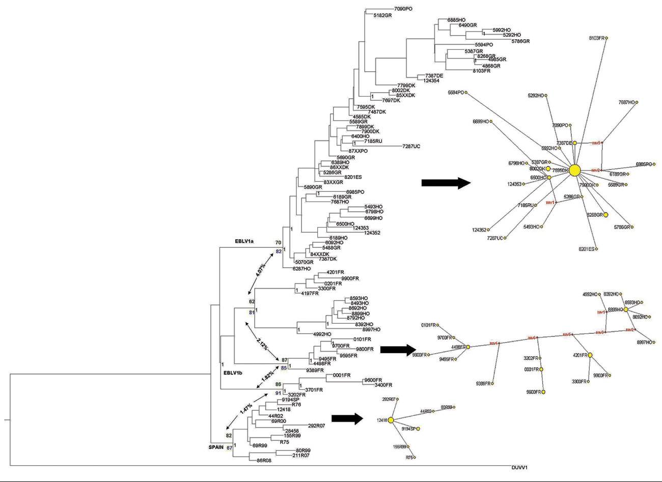
European bat lyssavirus 1 (EBLV-1) phylogenetic reconstruction based on the first 400 bp of the nucleoprotein gene. The tree was obtained by Bayesian inference run for 107 generations; trees were sampled every 100 generations. The first 25% of trees were excluded from the analysis as burn-in. Black numbers indicate posterior probabilities. Bootstrap supports after 1,000 replicates for each node are also shown for maximum-parsimony (green numbers) and maximum-likelihood (blue numbers) analyses. N

Figure 1. European bat lyssavirus 1 (EBLV-1) phylogenetic reconstruction based on the first 400 bp of the nucleoprotein gene. The tree was obtained by Bayesian inference run for 107 generations; trees were sampled every 100 generations. The first 25% of trees were excluded from the analysis as burn-in. Black numbers indicate posterior probabilities. Bootstrap supports after 1,000 replicates for each node are also shown for maximum-parsimony (green numbers) and maximum-likelihood (blue numbers) analyses. Net p-distance values (as percentages) between groups are indicated by arrows. A parsimony-based network is presented for each major lineage; sizes of yellow circles are proportional to the number of individuals sharing a given haplotype, and reconstructed haplotypes (median vectors) are shown in red. DUVV, Duvenhage virus.

Main Article

Page created: July 25, 2011
Page updated: July 25, 2011
Page reviewed: July 25, 2011
The conclusions, findings, and opinions expressed by authors contributing to this journal do not necessarily reflect the official position of the U.S. Department of Health and Human Services, the Public Health Service, the Centers for Disease Control and Prevention, or the authors' affiliated institutions. Use of trade names is for identification only and does not imply endorsement by any of the groups named above.
file_external