Coccidioides posadasii Infection in Bats, Brazil
Rossana de Aguiar Cordeiro

, Kylvia Rocha de Castro e Silva, Raimunda Sâmia Nogueira Brilhante, Francisco Bergson Pinheiro Moura, Naylê Francelino Holanda Duarte, Francisca Jakelyne de Farias Marques, Rebecca de Aguiar Cordeiro
, Renato Evando Moreira Filho, Roberto Wagner Bezerra de Araújo, Tereza de Jesus Pinheiro Gomes Bandeira, Marcos Fábio Gadelha Rocha, and José Júlio Costa Sidrim
Author affiliations: Universidade Federal do Ceará, Fortaleza-Ceará, Brazil (R.A. Cordeiro, K.R.C. Silva, R.S.N. Brilhante, F.J.F. Marques, R.A. Cordeiro, R.E. Moreira Filho, R.W.B. Araújo, T.J.P.G. Bandeira, M.F.G. Rocha, J.J.C. Sidrim); Instituto Federal de Educação, Ciência e Tecnologia, Ceará, Brazil (K.R.C. Silva); Universidade Estadual do Ceará, Fortaleza- Ceará (M.F.G. Rocha); Secretaria da Saúde do Estado do Ceará, Ceará (F.B.P. Moura, N.F.H. Duarte)
Main Article
Figure
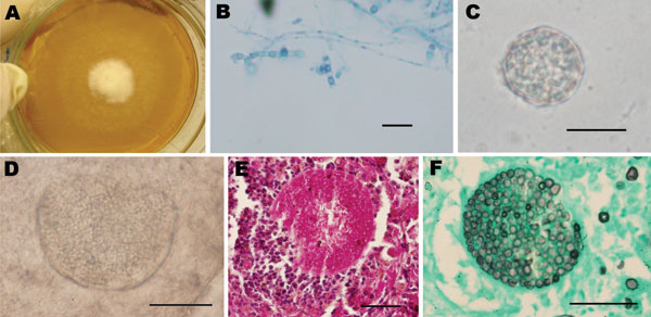
Figure. .
Coccidioidal structures obtained from a naturally infected Carollia perspicillata bat (upper images) and experimentally infected mice (lower images). A) Macroscopic aspect of Coccidioides posadasii culture recovered from homogenate of bat lungs. B) Microscopic view of C. posadasii culture from bat lungs showing hyaline hyphae with arthroconidia and disjunctor cells (lactophenol cotton blue staining). C) Mature spherule filled with endospores in lung tissue (10% KOH) of bat. D) Bursting spherule with endospores in mouse lung tissue (10% KOH). E) Histopathologic features of mouse lungs revealing parasitic coccidioidal forms by periodic acid-Schiff staining. F) Coccidioidal forms on mouse lungs shown by Grocott-Gomori methenamine-silver staining. Scale bars = 20 μm.
Main Article
Page created: March 15, 2012
Page updated: March 22, 2012
Page reviewed: March 22, 2012
The conclusions, findings, and opinions expressed by authors contributing to this journal do not necessarily reflect the official position of the U.S. Department of Health and Human Services, the Public Health Service, the Centers for Disease Control and Prevention, or the authors' affiliated institutions. Use of trade names is for identification only and does not imply endorsement by any of the groups named above.