Volume 19, Number 11—November 2013
Dispatch
Human Bocavirus in Patients with Encephalitis, Sri Lanka, 2009–2010
Abstract
We identified human bocavirus (HBoV) DNA by PCR in cerebrospinal fluid from adults and children with encephalitis in Sri Lanka. HBoV types 1, 2, and 3 were identified among these cases. Phylogenetic analysis of HBoV1 strain sequences found no subclustering with strains previously identified among encephalitis cases in Bangladesh.
Encephalitis is a serious infection causing high rates of illness and, in industrialized countries, has a case-fatality rate of 6.5%–12% (1,2). However, the situation in developing countries is largely unknown. Globally, the causes remain unrecognized in 60%–85% of encephalitis cases (1,2). Recently, human bocavirus (HBoV) has been implicated in causing life-threatening encephalitis in Bangladeshi children (3). In Sri Lanka, information about the causative agents of encephalitis is scarce. The aim of this study was to determine the occurrence of HBoV and other possible pathogens in children and adults with encephalitis admitted to a tertiary care hospital in Sri Lanka.
The study was conducted at Colombo North Teaching Hospital, Ragama, Sri Lanka, during July 2009–November 2010. A total of 233 patients (110 adolescents/adults >12 years of age and 123 children) were enrolled. Adolescents and adults were admitted to adult wards. Cerebrospinal fluid (CSF) samples were available from 191 patients. Criteria for enrolment were as follows: any combination of the triad of fever, headache, and vomiting, along with altered level of consciousness, seizures, focal neurologic deficits, altered behavior, and signs of meningeal irritation. Clinical and laboratory information was available for 164 patients. The male:female ratio for adolescents/adults was 1.3:1; ages ranged from 12 to 90 years (mean 42 years); For children, the male:female ratio was 0.7:1; ages ranged from 2 to 144 months (mean 48 months). The ethics committees of the University of Kelaniya and Oita University approved this study.
CSF samples were subjected to macroscopic examination, total and differential leukocyte counts, bacterial culture, Gram staining, and measurement of protein and glucose. Blood was cultured for bacteria and examined for total and differential leukocyte counts, erythrocyte sedimentation rates, and hemoglobin and C-reactive protein levels.
Classical encephalitis-causing pathogens (Table) and diarrheagenic viruses, such as HBoV, rotavirus, astrovirus, norovirus, parechovirus, and human adenovirus (HAdV), were determined in CSF by PCR (Technical Appendix) (3–5). Anti-
Nucleotide sequences of all amplicons were determined to confirm the PCR products, to distinguish genotypes, and to perform phylogenetic analysis (3). BLAST analysis (www.ncbi.nlm.nih.gov/blast) was used to identify the viruses and genotypes. Multiple sequence alignment was conducted by using ClustalW2 (www.ebi.ac.uk/clustalw). The phylogenetic analysis was done with a neighbor-joining tree by using MEGA5 (www.megasoftware.net). A bootstrap analysis of 1,000 replicates was performed to test the reliability of the branching pattern.
The causes of encephalitis were type 2 dengue virus in 1 (0.5%) patient, human echovirus (HEcoV) type 9 or 25 in 2 (1%), HBoV (Table) in 5 (3%), and HAdV 41 in 7 (4%): all were sole detections. None of the other viruses and no bacteria were detected. Samples positive for HBoV by primers designed from viral protein 1/2 also were positive by primers designed from nonstructural protein (NP) 1 gene. HEcoV was detected in 2- and 9-year-old children. HAdV 41 was not confined to children; ages of infected patients ranged from 13 months to 55 years. Of 81 CSF samples, anti-NMDAR encephalitis was detected in 2 (2%) adults (42 and 72 years of age). All patients in this study recovered and were discharged, except for one 13-month-old boy with HAdV 41 encephalitis who left the hospital against medical advice.
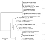
The severity of symptoms in the HBoV-positive patients did not differ from those of patients with other infections. None of the patients who had positive PCR results for HBoV1–3 had corresponding HBoV1–4 IgM or IgG in their CSF. Phylogenetic analysis (Figure) of the viral protein 1/2 gene showed that the Sri Lanka HBoV1 strains did not subcluster with encephalitis-associated Bangladesh strain, although they had 97%–98% nt identities. The Sri Lanka HBoV1 strains had 98%–99% nt identities among themselves and with other HBoV1 strains. The Sri Lanka HBoV2 strain was closely related to the Tunisia strain (96% nt identity). The Sri Lanka HBoV2 had 90%–91% nt identities with the Bangladeshi encephalitis-causing strains and 90%–96% nt identities with other HBoV2 strains. The Sri Lanka HBoV3 strain was closely associated with the cluster formed by viruses from the United Kingdom, Australia, Tunisia, and China and had 96%–97% nt identities with those strains. The sequence of NP1 gene is conserved and had 98%–100% nt identities among the Sri Lanka strains.
The study in Bangladesh suggested that HBoV-associated encephalitis might be restricted to malnourished children (3). However, our study demonstrates that HBoV also can be detected in well-nourished children and adults with encephalitis. How HBoV might trigger encephalitis is unclear. HBoV viremia has been documented, and the virus might therefore have the potential to cross the blood–brain barrier. The NP1 of HBoV inhibits interferon-β production by the host, suggesting evasion of the innate immune response during infection (8).
Unlike the Bangladesh study, where 2 of 4 encephalitis patients in whom HBoV was detected died (3), all patients in our study recovered. In addition to HBoV1 and HBoV2, we detected HBoV3 in a child with encephalitis, which to our knowledge, has not been reported as a cause of the disease. Although HBoV infections occur mainly in children, among the 5 Sri Lanka patients with HBoV encephalitis, 3 were adults or adolescents. None of the patients with HBoV encephalitis had HBoV IgM or IgG in their CSF, indicating how rapidly disease onset occurred and how little time the immune system had to respond. Generally, the specific seroprevalence rate of HBoV1 antibodies in infected persons is 59%, followed by HBoV2, 3, and 4 (34%, 15%, and 2%, respectively) (7).
Our detection rate of viruses as a cause of encephalitis was 7.5%, and adding anti-NMDAR encephalitis, the detection rate increased to 10%, which is similar to that of another study (9). Anti-NMDAR encephalitis is becoming a dominant cause of encephalitis in certain population (10); however, in Sri Lanka, it is 1%–4%, similar to other studies (11).
Dengue virus is the leading endemic cause of encephalitis in Brazil (12). This infection is also endemic to Sri Lanka and, before our study, dengue encephalitis was suspected but unconfirmed in the population. Enteroviruses frequently cause CNS infection, and the HEcoV 9 and 25 found here are known to cause encephalitis (13).
Among the HAdVs, serotype F is mainly responsible for gastroenteritis, whereas encephalitis is caused mainly by serotypes B, C, and D (14,15). The large number of HAdV 41 encephalitis cases indicates a unique epidemiology in Sri Lanka.
Herpes simplex and varicella-zoster viruses are implicated as the major causes of encephalitis. However, these viruses were not responsible for encephalitis in our study or in the studies in Bangladesh. HBoV is dominant in both Bangladesh and Sri Lanka. The limitation of our study is that causation could not be proven by the presence of HBoV antibody during infection or the absence of HBoV DNA in the CSF when recovered. The HBoV DNA detected in our study may represent persistent DNA from past infection; however, history of recent respiratory or diarrheal infection was absent. Future studies using quantitative PCR and serology are warranted to better establish the etiologic role of HBoV infection and encephalitis.
Mr Mori is a medical technologist and is enrolled in the PhD program in the Department of Microbiology, Faculty of Medicine, Oita University, Japan. His research interest is molecular epidemiology of viruses.
Acknowledgments
We thank Danushka Nawaratne, Maliduwa Liyanage Harshini, Nayomi Fonseka, Pradeepa Harshani, Ranjan Premaratne, Wathsala Hathagoda, Lasanthi Weerasooriya, Aruna Kulatunga, Nilmini Tissera, Manel Cooray, Lilanthi de Silva, Manel Fernando, and Thirughanam Sekar for their cooperation. We also thank Lea Hedman for performing enzyme immunoassay for HBoV1-4.
This study was supported in part by a Research Fund at the Discretion of the President, Oita University (grant no. 610000-N5010) to K.A., the University of Helsinki Research Funds to M.S-V., and the Grant for Comprehensive Research on Disability Health and Welfare (H24-002) from the Japan Ministry of Health and Labour Science Research to H.M.
References
- Granerod J, Ambrose HE, Davies NW, Clewley JP, Walsh AL, Morgan D, Causes of encephalitis and differences in their clinical presentations in England: a multicentre, population-based prospective study. Lancet Infect Dis. 2010;10:835–44. DOIPubMedGoogle Scholar
- Huppatz C, Durrheim DN, Levi C, Dalton C, Williams D, Clements MS, Etiology of encephalitis in Australia, 1990–2007. Emerg Infect Dis. 2009;15:1359–65 . DOIPubMedGoogle Scholar
- Mitui MT, Shahnawaz Bin Tabib SM, Matsumoto T, Khanam W, Ahmed S, Mori D, Detection of human bocavirus in the cerebrospinal fluid of children with encephalitis. Clin Infect Dis. 2012;54:964–7. DOIPubMedGoogle Scholar
- Han TH, Kim CH, Park SH, Chung JY, Hwang ES. Detection of human parechoviruses in children with gastroenteritis in South Korea. Arch Virol. 2011;156:1471–5. DOIPubMedGoogle Scholar
- Allander T, Tammi MT, Eriksson M, Bjerkner A, Tiveljung-Lindell A, Andersson B. Cloning of a human parvovirus by molecular screening of respiratory tract samples. Proc Natl Acad Sci U S A. 2005;102:12891–6. DOIPubMedGoogle Scholar
- Takano S, Takahashi Y, Kishi H, Taguchi Y, Takashima S, Tanaka K, Detection of autoantibody against extracellular epitopes of N-methyl-D-aspartate receptor by cell-based assay. Neurosci Res. 2011;71:294–302. DOIPubMedGoogle Scholar
- Kantola K, Hedman L, Arthur J, Alibeto A, Delwart E, Jartti T, Seroepidemiology of human bocaviruses 1–4. J Infect Dis. 2011;204:1403–12. DOIPubMedGoogle Scholar
- Zhang Z, Zheng Z, Luo H, Meng J, Li H, Li Q, Human bocavirus NP1 inhibits IFN-beta production by blocking association of IFN regulatory factor 3 with IFNB promoter. J Immunol. 2012;189:1144–53. DOIPubMedGoogle Scholar
- Dupuis M, Hull R, Wang H, Nattanmai S, Glasheen B, Fusco H, Molecular detection of viral causes of encephalitis and meningitis in New York State. J Med Virol. 2011;83:2172–81. DOIPubMedGoogle Scholar
- Gable MS, Sheriff H, Dalmau J, Tilley DH, Glaser CA. The frequency of autoimmune N-methyl-D-aspartate receptor encephalitis surpasses that of individual viral etiologies in young individuals enrolled in the California Encephalitis Project. Clin Infect Dis. 2012;54:899–904 . DOIPubMedGoogle Scholar
- Dalmau J, Lancaster E, Martinez-Hernandez E, Rosenfeld MR, Balice-Gordon R. Clinical experience and laboratory investigations in patients with anti-NMDAR encephalitis. Lancet Neurol. 2011;10:63–74. DOIPubMedGoogle Scholar
- Soares CN, Cabral-Castro MJ, Peralta JM, de Freitas MR, Zalis M, Puccioni-Sohler M. Review of the etiologies of viral meningitis and encephalitis in a dengue endemic region. J Neurol Sci. 2011;303:75–9. DOIPubMedGoogle Scholar
- Tavakoli NP, Wang H, Nattanmai S, Dupuis M, Fusco H, Hull R. Detection and typing of enteroviruses from CSF specimens from patients diagnosed with meningitis/encephalitis. J Clin Virol. 2008;43:207–11. DOIPubMedGoogle Scholar
- Frange P, Peffault de Latour R, Arnaud C, Boddaert N, Oualha M, Avettand-Fenoel V, Adenoviral infection presenting as an isolated central nervous system disease without detectable viremia in two children after stem cell transplantation. J Clin Microbiol. 2011;49:2361–4. DOIPubMedGoogle Scholar
- Straussberg R, Harel L, Levy Y, Amir J. A syndrome of transient encephalopathy associated with adenovirus infection. Pediatrics. 2001;107:E69. DOIPubMedGoogle Scholar
Figure
Table
Cite This ArticleTable of Contents – Volume 19, Number 11—November 2013
| EID Search Options |
|---|
|
|
|
|
|
|

Please use the form below to submit correspondence to the authors or contact them at the following address:
Kamruddin Ahmed, Research Promotion Institute, Oita University, Yufu 879-5593, Oita, Japan
Top