Volume 19, Number 3—March 2013
Letter
Melioidosis and Hairy Cell Leukemia in 2 Travelers Returning from Thailand
Benjamin Rossi1, Loïc Epelboin1 , Stéphane Jauréguiberry, Maryline Lecso, Damien Roos-Weil, Jean Gabarre, Philippe A. Grenier, François Bricaire, and Eric Caumes
Figure
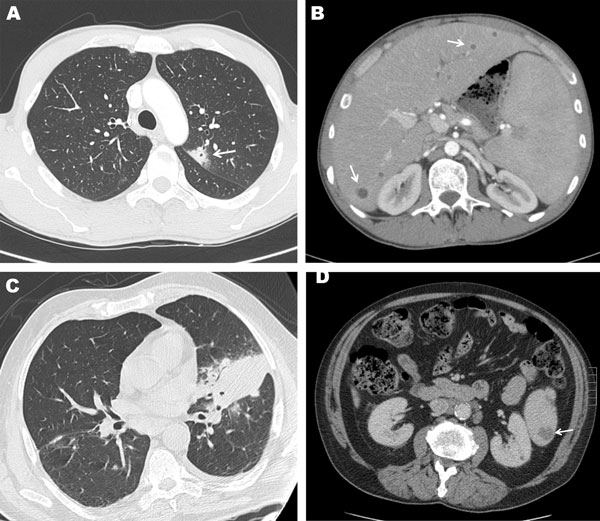
Figure. . . Computed tomography (CT) images of the chest and abdomen of case-patient 1 showing A) a subpleural nodular and cavitary lesion (arrow) in the left upper lobe of the lung and B) multiple small round liver abscesses, seen as multiple foci of ill-defined areas of hypoattenuation (arrows), and enlargement of the spleen. CT images of the chest and abdomen of case-patient 2 showing C) a focal area of parenchymal consolidation in the left lung associated with an ipsilateral mild pleural effusion and D) and a spleen abscess (arrow).
1These authors contributed equally to this article.
Page created: February 12, 2013
Page updated: February 12, 2013
Page reviewed: February 12, 2013
The conclusions, findings, and opinions expressed by authors contributing to this journal do not necessarily reflect the official position of the U.S. Department of Health and Human Services, the Public Health Service, the Centers for Disease Control and Prevention, or the authors' affiliated institutions. Use of trade names is for identification only and does not imply endorsement by any of the groups named above.