Volume 19, Number 9—September 2013
Dispatch
Human Parainfluenza Virus Type 3 in Wild Nonhuman Primates, Zambia
Michihito Sasaki, Akihiro Ishii, Yasuko Orba, Yuka Thomas, Bernard M. Hang’ombe, Ladslav Moonga, Aaron S. Mweene, Hirohito Ogawa, Ichiro Nakamura, Takashi Kimura, and Hirofumi Sawa
Figure 2
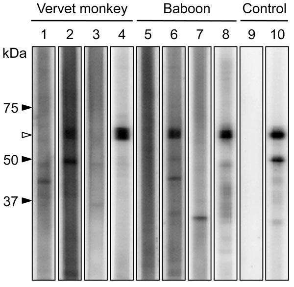
Figure 2. . Western blot analysis of purified recombinant N protein from human parainfluenza virus type 3. Western blot analysis was performed by using serum specimens from vervet monkeys (lanes 1–4) and baboons (lanes 5–8) in the Mfuwe (lanes 1, 2, 5, 6) and Livingstone (lanes 3, 4, 7, 8) regions. Results of representative antibody-negative (lanes 1, 3, 5, 7) and antibody-positive (lanes 2, 4, 6, 8) samples are shown. Mock antibody (lane 9) and HPIV monoclonal antibody (lane 10) were used as negative and positive controls, respectively. The putative molecular mass of the recombinant N protein (white arrowhead) is 59 kDa.
Page created: August 20, 2013
Page updated: August 20, 2013
Page reviewed: August 20, 2013
The conclusions, findings, and opinions expressed by authors contributing to this journal do not necessarily reflect the official position of the U.S. Department of Health and Human Services, the Public Health Service, the Centers for Disease Control and Prevention, or the authors' affiliated institutions. Use of trade names is for identification only and does not imply endorsement by any of the groups named above.