Skip directly to site content Skip directly to page options Skip directly to A-Z link Skip directly to A-Z link Skip directly to A-Z link
Volume 20, Number 1—January 2014
Research

Molecular Barriers to Zoonotic Transmission of Prions

Marcelo A. Barria, Aru Balachandran, Masanori Morita, Tetsuyuki Kitamoto, Rona Barron, Jean Manson, Richard Knight, James W. Ironside, and Mark W. HeadComments to Author 
Author affiliations: The University of Edinburgh, Edinburgh, Scotland, UK (M.A. Barria, R. Knight, J.W. Ironside, M.W Head); Canadian Food Inspection Agency, Ottawa, Ontario, Canada (A. Balachandran); Japan Blood Products Organization, Kobe, Japan (M. Morita); Tohoku University Graduate School of Medicine, Sendai, Japan (T. Kitamoto); University of Edinburgh, Easter Bush, Scotland, UK (R. Barron, J. Manson)

Main Article

Figure 1

Determination of total PrP and PrPres level in animal tissues. To characterize the PrP expression levels (total PrP), brain homogenates were analyzed by Western blot without digestion with PK. Nineteen microliters of each 10% wt/vol homogenate was loaded in each lane (A). To detect the PrPres in the samples, PK digestion (50 μg/mL) was performed to remove PrPC, and the samples were then reanalyzed (B). The atypical scrapie and matched normal control animal samples were further analyzed by Wester

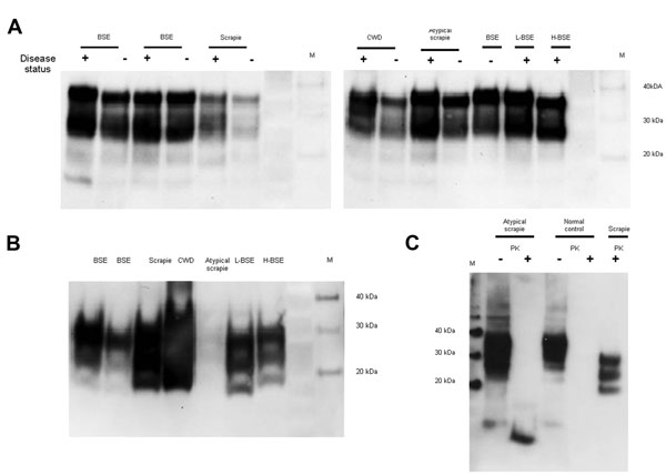
Figure 1. . Determination of total PrP and PrPres level in animal tissues. To characterize the PrP expression levels (total PrP), brain homogenates were analyzed by Western blot without digestion with PK. Nineteen microliters of each 10% wt/vol homogenate was loaded in each lane (A). To detect the PrPres in the samples, PK digestion (50 μg/mL) was performed to remove PrPC, and the samples were then reanalyzed (B). The atypical scrapie and matched normal control animal samples were further analyzed by Western blot both with (+) and without (–) prior PK digestion (C) by comparing 3 μL of undigested homogenates with 100 μL of the PK-digested sample concentrated by centrifugation. Five microliters of a PK-digested classical scrapie brain homogenate was analyzed in parallel for comparison. The detection antibody was 6H4 in (A) and (B) and 9A2 in (C). PrP, prion protein; PrPres, protease-resistant PrP; PK, proteinase K; PrPc, normal cellular PrP; M, molecular marker; BSE, bovine spongiform encephalopathy; +, animal prion disease sample; –, matched normal animal control sample; CWD, chronic wasting disease; L-BSE, L-type BSE; H-BSE, H-type BSE.

Main Article

Page created: January 03, 2014
Page updated: January 03, 2014
Page reviewed: January 03, 2014
The conclusions, findings, and opinions expressed by authors contributing to this journal do not necessarily reflect the official position of the U.S. Department of Health and Human Services, the Public Health Service, the Centers for Disease Control and Prevention, or the authors' affiliated institutions. Use of trade names is for identification only and does not imply endorsement by any of the groups named above.
file_external