Volume 22, Number 2—February 2016
Dispatch
Frequency and Distribution of Rickettsiae, Borreliae, and Ehrlichiae Detected in Human-Parasitizing Ticks, Texas, USA
Cite This Article
Citation for Media
Abstract
To describe the presence and distribution of tickborne bacteria and their vectors in Texas, USA, we screened ticks collected from humans during 2008–2014 for Rickettsia, Borrelia, and Ehrlichia spp. Thirteen tick species were identified, and 23% of ticks carried bacterial DNA from at least 1 of the 3 genera tested.
Ticks are vectors for a variety of microorganisms, many of which are known agents of zoonotic disease. Although much current research is focused on areas where these diseases are common, it is crucial to collect data from areas with fewer diagnoses of tickborne illness. In Texas, USA, tickborne diseases caused by Rickettsia, Borrelia, and Ehrlichia bacteria are diagnosed less frequently than in some areas of the United States (1); however, those agents have been documented to occur (2), and many medically relevant tick species, capable of carrying and transmitting these pathogens, are established in various geographic areas of Texas (1). Long-term surveillance data encompassing consecutive seasons and a wide geographic range are necessary to ascertain disease transmission risks associated temporally or geographically with established or emerging tickborne pathogens and their vectors. The University of North Texas Health Science Center Tick-Borne Disease Research Laboratory (UNTHSC-TBDL), the primary tick-testing facility for Texas Department of State Health Services Zoonosis Control (TX DSHS), receives ticks continually throughout the year. The data collected from this testing provide an assessment of the prevalence of tick species and associated tickborne bacterial agents collected in Texas.
From October 1, 2008, through September 30, 2014, ticks removed from humans were sent by TX DSHS to UNTHSC-TBDL, where they were tested by using PCR-based methods, then underwent by DNA sequence analysis to determine the presence of Rickettsia, Borrelia, and Ehrlichia spp. Morphologic identification of tick species was implemented by entomologists at TX DSHS. Ticks that could not be classified morphologically were identified at UNTHSC-TBDL by sequencing mitochondrial 16S rDNA (data not shown).
Each tick was sent to UNTHSC-TBDL in an individual collection tube. Upon arrival, ticks were processed according to the laboratory’s standard protocol, as described by Williamson et al. (2). After bead pulverization, we extracted DNA using the E.Z.N.A. Mollusc DNA Isolation Kit (Omega Bio-Tek, Norcross, GA, USA) following the manufacturer’s protocol.
DNA from each specimen was screened in duplicate by PCR for Rickettsia, Borrelia, and Ehrlichia spp. as previously described (2) by using primers listed in Table 1. PCR products were evaluated, and presumptive-positive amplicons were purified for sequencing (2). Cycle sequencing reactions were performed in both directions by using BigDye Terminator version 3.1 chemistry (Life Technologies, Carlsbad, CA, USA). Dideoxy chain termination products were detected electrophoretically on an ABI 310 or 3130xL Genetic Analyzer (Life Technologies). Sequence analysis was performed by using Sequencher version 4.8/5.0 (GeneCodes, Ann Arbor, MI, USA). Analyzed sequences were compared with reference data in GenBank (http://blast.ncbi.nlm.nih.gov/). Sequences were submitted to GenBank under accession nos. KP861333–KP861347.
The TX DSHS submitted 1,112 ticks to UNTHSC-TBDL during October 1, 2008–September 30, 2014, of which 1,062 originated in Texas. Thirteen tick species were identified; most were Amblyomma americanum (55.7%), followed by Dermacentor variabilis (15.0%), Rhipicephalus sanguineus (13.0%), Ixodes scapularis (5.6%), A. maculatum (5.4%), and A. cajennense (2.9%). Approximately 23.3% of ticks originating in Texas tested positive for DNA from Rickettsia, Borrelia, or Ehrlichia bacteria (Table 2; Technical Appendix Table). Of these bacteria, most belonged to spotted fever group rickettsiae (SFGR); A. americanum was the most common tick species found to carry an SFGR agent. The most frequent SFGR sequences detected demonstrated 100% identity to Candidatus Rickettsia amblyommii rompA (GenBank accession no. EF194096). Candidatus R. amblyommii was detected in both A. americanum and A. cajennense ticks and showed prevalence rates of 30.3% and 32.3%, respectively. The second most common SFGR rompA sequences were 100% homologous to the previously termed rickettsial I. scapularis endosymbiont, which has been officially named R. buchneri (accession no. KP172259) (9). Five A. maculatum specimens contained DNA sequences identical to R. parkeri rompA (accession no. KC003476). Sequences that shared 100% similarity to 1 specific R. rhipicephali isolate (accession no. U43803) and 99% similarity to other R. rhipicephali rompA isolates (accession nos. EU109175–EU109178) were obtained from 4 D. variabilis ticks. Sequences isolated from 2 D. andersoni ticks were identical to R. peacockii rompA and rompB (accession nos. FM883671 and CP001227, respectively). Tick species was confirmed by sequencing mitochondrial 16S rDNA. Sequences from both specimens aligned 99% with D. andersoni (accession no. EU711343) and 94% with D. variabilis (accession no. L34300). D. andersoni is not known to inhabit Texas (1,10), so this finding could suggest a novel geographic association.
The total prevalence of borreliae detected was 1.1%. DNA sequences sharing 100% identity to B. lonestari were found in 8 A. americanum ticks (1.4%). As seen by Stromdahl et al., the B. lonestari isolates matching sequences in this study depended on the insertion or deletion of a nucleotide triplet, AAG (11). Sequences from 7 tick samples matched 100% with B. lonestari flaB isolates containing the additional triplet (accession no. AY850063), and 1 sequence was identical to B. lonestari flaB isolates lacking the triplet (accession no. AY850064). Of the 8 A. americanum ticks from which the B. lonestari sequences were obtained, 6 were co-infected with Candidatus R. amblyommii. DNA extracts from 1 I. scapularis tick contained a sequence consistent with B. burgdorferi sensu stricto (s.s.) and was co-infected with R. buchneri. The flaB sequence matched 100% to (accession no. CP002228), and 99% to (accession no. CP009656) B. burgdorferi s.s. reference sequences. The Borrelia 16S rDNA sequence showed 100% identity to (accession no. CP009656) and differed by 1 single nucleotide polymorphism from (accession no. CP002228) B. burgdorferi s.s. reference sequences. A flaB gene sequence from 1 D. variabilis tick shared 100% identity with Candidatus B. texasensis (accession no. AF264901). Samples from 2 A. maculatum ticks showed flaB sequences matching 90% identity values to B. turcica (accession no. AB109243), a reptilian Borrelia sp. Those flaB sequences were identical to a novel Borrelia sp. (accession no. KF395230) previously found in A. maculatum ticks in Mississippi and known to share a phylogenetic clade with B. turcica (12). Borrelia 16S rDNA primers produced nonspecific amplification with these 2 samples.
Figure
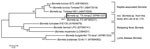
Phylogenetic analysis was performed by using MEGA version 5.1 (http://www.megasoftware.net) using GenBank reference sequences to examine relationships between the Borrelia sp. from this study, B. turcica, and both Lyme disease–associated and relapsing fever borreliae (Figure). The results supported findings by Lee et al. that the novel Borrelia sp. flaB sequences were more closely related to the reptilian Borrelia than the other 2 Borrelia groups (12).
Two A. americanum ticks contained DNA sharing 100% identity with Ehrlichia chaffeensis dsb (accession no. CP000236). One of these ticks was co-infected with Candidatus R. amblyommii. Prevalence of E. chaffeensis in the A. americanum specimens tested was 0.34%. In addition, 2 of 42 A. maculatum ticks tested for the emerging pathogen Panola Mountain Ehrlichia sp. (PME) (7) each produced a map1 sequence that was 100% homologous to 2 separate PME reference sequences (accession nos. EU272356, EU272358). These sequences differed from each other by 1 single nucleotide polymorphism. This finding represents a novel association, as A. americanum is the known vector for PME (7). A subset of 141 A. americanum ticks was also tested for PME, with negative results.
Frequency of tickborne zoonoses in Texas remains low compared with some regions of the United States. We report the detection of known pathogens along with bacteria of unknown pathogenicity in human-parasitizing ticks commonly found in Texas. Our findings underscore the importance of better characterization and continued surveillance of the frequency and distribution of tick species and the bacterial agents they carry. Continued monitoring in low-risk areas provides data regarding the presence of potential emerging pathogens and vectors not yet commonly identified, which could pose unidentified threats to public health.
Ms. Mitchell is a tick-borne disease analyst at the University of North Texas Health Science Center Tick-Borne Disease Research Laboratory in the Center for Biosafety and Biosecurity. Her current research focuses on detection, identification, and characterization of pathogens and potential emerging agents of human disease.
Acknowledgments
We thank Rhonda Roby for advice regarding this work. We also thank Bonny Mayes, Jim Schuermann, Dave Florin, and staff at Texas DSHS Zoonosis Control for collection and taxonomic identification of tick samples.
This project was financially supported by the State of Texas.
References
- Centers for Disease Control and Prevention, National Center for Emerging and Zoonotic Infectious Diseases, Division of Vector-Borne Diseases. Geographic distribution of ticks that bite humans [cited 2015 Aug 20]. http://www.cdc.gov/ticks/geographic_distribution.html
- Williamson PC, Billingsley PM, Teltow GJ, Seals JP, Turnbough MA, Atkinson SF. Borrelia, Ehrlichia, and Rickettsia spp. in ticks removed from persons, Texas, USA. Emerg Infect Dis. 2010;16:441–6. DOIPubMedGoogle Scholar
- Barbour AG, Maupin GO, Teltow GJ, Carter CJ, Piesman J. Identification of an uncultivable Borrelia species in the hard tick Amblyomma americanum: possible agent of a Lyme disease-like illness. J Infect Dis. 1996;173:403–9. DOIPubMedGoogle Scholar
- Regnery RL, Spruill CL, Plikaytis BD. Genotypic identification of rickettsiae and estimation of intraspecies sequence divergence for portions of two rickettsial genes. J Bacteriol. 1991;173:1576–89 .PubMedGoogle Scholar
- Eremeeva M, Yu X, Raoult D. Differentiation among spotted fever group rickettsiae species by analysis of restriction fragment length polymorphism of PCR-amplified DNA. J Clin Microbiol. 1994;32:803–10 .PubMedGoogle Scholar
- Doyle CK, Labruna MB, Breitschwerdt EB, Tang YW, Corstvet RE, Hegarty BC, Detection of medically important Ehrlichia by quantitative multicolor TaqMan real-time polymerase chain reaction of the dsb gene. J Mol Diagn. 2005;7:504–10. DOIPubMedGoogle Scholar
- Loftis AD, Mixson TR, Stromdahl EY, Yabsley MJ, Garrison LE, Williamson PC, Geographic distribution and genetic diversity of the Erhlichia sp. from Panola Mountain in Amblyomma americanum. BMC Infect Dis. 2008;8:54. DOIPubMedGoogle Scholar
- Black WC IV, Piesman J. Phylogeny of hard- and soft-tick taxa (Acari: Ixodida) based on mitochondrial 16S rDNA sequences. Proc Natl Acad Sci U S A. 1994;91:10034–8. DOIPubMedGoogle Scholar
- Kurtti TJ, Felsheim RF, Burkhardt NY, Oliver JD, Heu CC, Munderloh UG. Rickettsia buchneri sp. nov., a rickettsial endosymbiont of the blacklegged tick Ixodes scapularis. Int J Syst Evol Microbiol. 2015;65:965–70. DOIPubMedGoogle Scholar
- James AM, Freier JE, Keirans JE, Durden LA, Mertins JW, Schlater JL. Distribution, seasonality, and hosts of the Rocky Mountain wood tick in the United States. J Med Entomol. 2006;43:17–24. DOIPubMedGoogle Scholar
- Stromdahl EY, Williamson PC, Kollars TM Jr, Evans SR, Barry RK, Vince MA, Evidence of Borrelia lonestari DNA in Amblyomma americanum (Acari: Ixodidae) removed from humans. J Clin Microbiol. 2003;41:5557–62. DOIPubMedGoogle Scholar
- Lee JK, Smith WC, McIntosh C, Ferrari FG, Moore-Henderson B, Varela-Stokes A. Detection of a Borrelia species in questing Gulf Coast ticks, Amblyomma maculatum. Ticks Tick Borne Dis. 2014;5:449–52.
Figure
Tables
Cite This ArticleTable of Contents – Volume 22, Number 2—February 2016
| EID Search Options |
|---|
|
|
|
|
|
|
Please use the form below to submit correspondence to the authors or contact them at the following address:
Michael S. Allen, University of North Texas Health Science Center, Department of Molecular and Medical Genetics, 3500 Camp Bowie Blvd, Fort Worth, TX 76107, USA
Top