Sneathia amnii and Maternal Chorioamnionitis and Stillbirth, Mozambique
Pio Vitorino
1, Rosauro Varo
1
, Paola Castillo, Juan Carlos Hurtado, Fabiola Fernandes, Ana Marta Valente, Rita Mabunda, Sibone Mocumbi, Joy M. Gary, Tiffany G. Jenkinson, Inacio Mandomando, Dianna M. Blau, Robert F. Breiman, and Quique Bassat
Author affiliations: Centro de Investigação em Saúde de Manhiça, Maputo, Mozambique (P. Vitorino, R. Varo, A.M. Valente, R. Mabunda, I. Mandomando, Q. Bassat); ISGlobal Hospital Clinic–Universitat de Barcelona, Barcelona, Spain (R. Varo, P. Castillo, J.C. Hurtado, A.M. Valente, Q. Bassat); Hospital Clínic, Barcelona (P. Castillo, J.C. Hurtado); Hospital Central de Maputo, Maputo (F. Fernandes, S. Mocumbi); Universidade Eduardo Mondlane, Maputo (F. Fernandes, S. Mocumbi); Centers for Disease Control and Prevention, Atlanta, Georgia, USA (J.M. Gary, T.G. Jenkinson, D.M. Blau); Emory Global Health Institute, Atlanta (R.F. Breiman); Institució Catalana de Recerca i Estudis Avançats (ICREA), Barcelona (Q. Bassat); Hospital Sant Joan de Déu, Barcelona (Q. Bassat); Consorcio de Investigación Biomédica en Red de Epidemiología y Salud Pública (CIBERESP),; Madrid, Spain (Q. Bassat)
Main Article
Figure
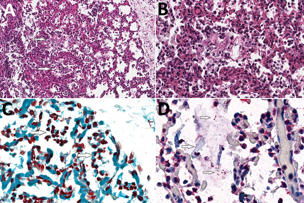
Figure. Histologic evidence of amniotic fluid aspiration, bronchopneumonia, and intraalveolar gram-negative coccobacilli in the lung of a stillborn infant, Mozambique. A) Hematoxylin and eosin stain of lung tissue showing acute inflammation within alveoli (bronchopneumonia, upper arrow) and moderate numbers of aspirated squames (lower arrow), consistent with intrauterine fetal distress and associated aspiration of amniotic fluid. Original magnification ×20. B) Higher magnification of panel A tissue showing acute inflammation within alveoli (bronchopneumonia) and a clump of aspirated squames. Original magnification ×40. C) Gram stain of lung showing multiple small, gram-negative coccobacilli mixed with acute inflammation within alveoli (arrows indicate regions with bacteria). Original magnification ×63. D) Polybacterial immunohistochemical assay of lung tissue targeting multiple bacteria highlights the coccobacilli within alveoli (top and bottom arrows). Aspirated squames are also present (middle arrow). Original magnification ×63.
Main Article
Page created: July 17, 2019
Page updated: July 17, 2019
Page reviewed: July 17, 2019
The conclusions, findings, and opinions expressed by authors contributing to this journal do not necessarily reflect the official position of the U.S. Department of Health and Human Services, the Public Health Service, the Centers for Disease Control and Prevention, or the authors' affiliated institutions. Use of trade names is for identification only and does not imply endorsement by any of the groups named above.