Volume 3, Number 2—June 1997
Synopsis
Rhodococcus equi and Arcanobacterium haemolyticum: Two "Coryneform" Bacteria Increasingly Recognized as Agents of Human Infection
Regina Linder
Figure 1
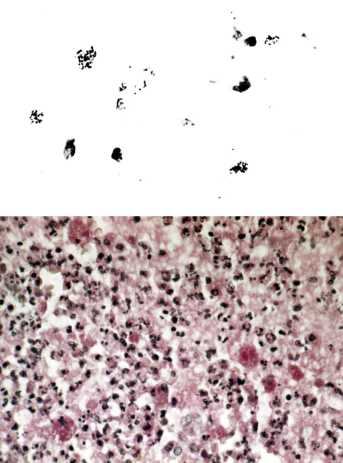
Figure 1. A. Bronchial tissue Gram stain showing intrahistiocytic coccobacillary forms of Rhodococcus equi. Original magnification, x 1,000. B.Open lung biopsy showing coalescent microabscesses with numerous histiocytes containing Rhodococcus equi organisms. PAS stain. Original magnification x 250. Figure provided by Dr. Margie Scott, Vanderbilt University Medical Center.
Page created: December 21, 2010
Page updated: December 21, 2010
Page reviewed: December 21, 2010
The conclusions, findings, and opinions expressed by authors contributing to this journal do not necessarily reflect the official position of the U.S. Department of Health and Human Services, the Public Health Service, the Centers for Disease Control and Prevention, or the authors' affiliated institutions. Use of trade names is for identification only and does not imply endorsement by any of the groups named above.