Volume 6, Number 6—December 2000
Dispatch
Genotypic Analysis of Multidrug-Resistant Salmonella enterica Serovar Typhi, Kenya
Cite This Article
Citation for Media
Abstract
We report the emergence in Kenya during 1997-1999 of typhoid fever due to Salmonella enterica serovar Typhi resistant to ampicillin, tetracycline, chloramphenicol, streptomycin, and cotrimoxazole. Genotyping by pulsed-field gel electrophoresis of XbaI-digested chromosomal DNA yielded a single cluster. The multidrug-resistant S. Typhi were related to earlier drug-susceptible isolates but were unrelated to multidrug-resistant isolates from Asia.
Salmonella enterica serovar Typhi causes approximately 10 million cases of typhoid that result in 600,000 deaths each year, mostly in developing countries (1). The antibiotics that form the mainstay of therapy in developing countries are chloramphenicol, ampicillin, and cotrimoxazole. Multidrug-resistant (MDR) strains of S. Typhi (resistant to all the above antimicrobial drugs) have caused outbreaks in the Indian subcontinent, Southeast Asia, and the Middle East since 1987 (2). Genetic studies have shown that resistance is encoded on an HI1 incompatibility plasmid and is transferable (3). MDR S. Typhi has not caused problems in Africa, except in South Africa (4), nor in South and Central America (5), and most isolates have remained fully susceptible. During 1997-1999, a number of isolates of MDR S. Typhi were identified from patients with typhoid in Nairobi, Kenya. We have examined their genotypic relationship to each other, to sensitive strains from Nairobi, and to MDR S. Typhi from Southeast Asia.
We analyzed isolates of S. Typhi obtained in the Kenyatta National Hospital from blood cultures of 16 adults with typhoid from 1988 to 1993; from 22 cultures of 19 adults and 3 children from 1997 to 1999; and from 17 representative MDR S. Typhi strains collected from 1990 to 1995 (6) from Pakistan (7), Hong Kong (4), Bangladesh (3), Kuwait (1), and India (1). We did not have access to isolates from 1994 to 1996, when no active surveillance was conducted. MICs of ampicillin, coamoxyclav, cefuroxime, cotrimoxazole, chloramphenicol, gentamicin, streptomycin, tetracycline, nalidixic acid, and ciprofloxacin were determined by the E-test method (AB Biodisk, Solna, Sweden).
Macrorestricted (using XbaI) chromosomal DNA from the S. Typhi isolates was separated by pulsed-field gel electrophoresis (PFGE) with a CHEF DRII system (Bio-Rad Labs, Richmond, VA). The gels were stained with ethidium bromide and photographed on an ultraviolet transilluminator. Banding patterns were compared (8), and dendrograms of relatedness were constructed by data clustering using the unweighted pair grouping arithmetic averaging method (Molecular Fingerprinting Program version 1.4.1, BioRad). Conjugation experiments, plasmid extraction and electrophoresis, and incompatibility grouping were performed as described (7).
All 16 S. Typhi isolates from 1988-1993 were fully sensitive to all the drugs tested (MIC 0.012-0.016 mg/L for ciprofloxacin to 1-3 mg/L for chloramphenicol). In contrast, 18 (82%) of the 22 S. Typhi from 1997-1999 were resistant to ampicillin, tetracycline, and chloramphenicol (MICs all > 32 mg/L), as were the 17 isolates from Asia. The first two MDR S. Typhi from Kenya were detected from blood cultures from two adults in March 1997. Active surveillance is ongoing, and multidrug resistance is detected in approximately 65% of all S. Typhi isolates to date. As we did not have access to isolates from 1994-1996, we cannot be certain that MDR S. Typhi did not emerge earlier than 1997.
All the Kenyan MDR S. Typhi isolates came from indigenous patients with no known history of recent travel outside the country. The Kenyan MDR isolates remained sensitive to co-amoxyclav, cefuroxime, gentamicin, nalidixic acid, and ciprofloxacin. The Kenyan MDR S. Typhi all transferred their full resistance phenotype to Escherichia coli K12 on 98-100 MDa plasmids of inc HI1 (or inc HI1 cross-reacting with inc FIIA).
Figure 1
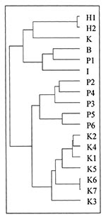
Figure 1. Dendogram showing genetic relatedness of Salmonella Typhi from Kenya and Asia. H1 and H2: MDR S. Typhi from Hong Kong; K: MDR S. Typhi from Kuwait; B: MDR S. Typhi from...
Figure 2
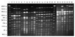
Figure 2. XbaI restriction endonuclease fragment patterns of representative Salmonella Typhi isolates from various countries. Lanes 1 and 19, molecular size standard; Lane 2, B1 from Bangladesh; Lane 3, I1 from India; Lanes...
All 22 MDR and 16 sensitive S. Typhi were analyzed by PFGE. As all 22 MDR isolates were similar by PFGE, only two representative strains were selected for further analysis. In addition, 5 representative sensitive S. Typhi and 11 representative MDR strains from Asia were analyzed for similarity by using dendograms. The sensitive S. Typhi (1987-1992) had a number of different genotypes. The MDR S. Typhi were identical, but differed from the sensitive isolates by more than seven bands, indicating they were different strains. However, on the dendogram comparing MDR S. Typhi from Asia and the S. Typhi from Kenya (both MDR and sensitive), the Kenyan isolates formed one cluster, with the nearest (but genotypically quite distinct) other cluster being the Pakistani MDR S. Typhi (Figure 1 and Figure 2).
The emergence of an MDR S. Typhi strain in Kenya is of concern because resistance to first-line antibiotics that are also commonly used for treatment of other bacterial infections in hospitals may pose a major challenge to health care. Although these newly emerged MDR S. Typhi are sensitive to nalidixic acid and ciprofloxacin, their MICs are five and ten times higher, respectively, than those of the sensitive S. Typhi from 1988-1993. Although fluoroquinolones are not widely available in Kenya, they may be needed to treat MDR S. Typhi, and resistance will lead to problems with treatment, as in Asia (9). Multidrug-resistant S. Typhi isolates from Kenya produced an indistinguishable PFGE pattern that was related to those of sensitive strains but unrelated to those of MDR S. Typhi from Asia. This finding implies that the Kenyan MDR S. Typhi are most likely to have arisen from sensitive isolates by acquisition of resistance plasmids from antibiotic-resistant enteric bacteria. Plasmids of incompatibility group HI1 are those most frequently found in S. Typhi, but we did not detect them in any of our nontyphoidal salmonellae with the same plasmid-encoded resistance (7).
We observed the emergence of S. Typhi resistant to all first-line drugs used for treatment of typhoid in Kenya and in many other African countries. Laboratories in Kenya should perform surveillance by routinely testing S. Typhi for susceptibility to first-line treatment drugs and to nalidixic acid to detect quinolone resistance. Effective surveillance for this newly emerged MDR S. Typhi in Africa and other developing regions of the world where MDR S. Typhi has not yet emerged would ensure prompt diagnosis, susceptibility testing, and appropriate antimicrobial chemotherapy.
Dr. Kariuki is a senior research officer at the Centre for Microbiology Research, Kenya Medical Research Institute. His research interests are in epidemiologic and genetic characterization of enteric bacteria and antibiotic resistance.
Acknowledgments
We thank Davy Koech, Director, Kenya Medical Research Institute, for support of publication of this work.
Financial support was provided by the Wellcome Trust.
References
- Pang T, Levine MM, Ivanoff B, Wain J, Finlay BB. Typhoid fever: important issues still remain. Trends Microbiol. 1998;6:131–3. DOIPubMedGoogle Scholar
- Mirza SH, Beeching NJ, Hart CA. Multidrug resistant typhoid: a global problem. J Med Microbiol. 1996;44:317–9. DOIPubMedGoogle Scholar
- Shanahan PMA, Jesudason MV, Thomson CJ, Amyes SGB. Molecular analysis of and identification of antibiotic resistance genes in clinical isolates of Salmonella typhi from India. J Clin Microbiol. 1998;36:1595–600.PubMedGoogle Scholar
- Coovadia YM, Gathiram V, Bhamjee A, Garratt RM, Mlisana K, Pillay N, An outbreak of multiresistant Salmonella typhi in South Africa. Q J Med. 1992;82:91–100.PubMedGoogle Scholar
- Olarte J, Galindo E. resistant to chloramphenicol, ampicillin, and other antimicrobial agents: strains isolated during an extensive typhoid fever epidemic in Mexico. Antimicrob Agents Chemother. 1973;4:597–601.PubMedGoogle Scholar
- Mirza S, Kariuki S, Mamun KZ, Beeching NJ, Hart CA. Analysis of plasmid and chromosomal DNA of multidrug-resistant Salmonella enterica Serovar Typhi from Asia. J Clin Microbiol. 2000;38:1449–52.PubMedGoogle Scholar
- Kariuki S, Gilks C, Corkill J, Kimari J, Benea A, Waiyaki P, Multidrug resistant non-typhi Salmonellae in Kenya. J Antimicrob Chemother. 1996;38:425–34. DOIPubMedGoogle Scholar
- Tenover FC, Arbeit RD, Goering RV, Mickelsen PA, Murray BE, Persing DH, Interpreting chromosomal DNA restriction patterns produced by pulsed-field gel electrophoresis: criteria for bacterial strain typing. J Clin Microbiol. 1995;33:2233–9.PubMedGoogle Scholar
- Murdoch DA, Banatvaia NA, Bone A, Shoismatulloev BI, Ward LR, Threlfall JE. Epidemic of ciprofloxacin-resistant Salmonella typhi in Tajikistan. Lancet. 1998;351:339. DOIPubMedGoogle Scholar
Figures
Cite This ArticleTable of Contents – Volume 6, Number 6—December 2000
| EID Search Options |
|---|
|
|
|
|
|
|
Please use the form below to submit correspondence to the authors or contact them at the following address:
Sam Kariuki, Centre for Microbiology Research, Kenya Medical Research Institute, P.O. Box 43640, Nairobi, Kenya; Fax: 254-2-711673
Top