Volume 7, Number 1—February 2001
Dispatch
Shigella spp. Surveillance in Indonesia: the Emergence or Reemergence of S. dysenteriae
Cite This Article
Citation for Media
Abstract
From June 1998 through November 1999, Shigella were isolated in 5% of samples from 3,848 children and adults with severe diarrheal illness in hospitals throughout Indonesia. S. dysenteriae has reemerged in Bali, Kalimantan, and Batam and was detected in Jakarta after a hiatus of 15 years.
Shigella spp. cause acute, debilitating diarrheal disease in humans (particularly young children) worldwide (1). In developing countries, where affected populations are immunologically compromised by poor nutrition and background infections, deaths attributed to shigellosis are common. Four Shigella species are recognized as pathogenic to humans: S. sonnei, S. boydii, S. flexneri, and S. dysenteriae. Both S. sonnei and S. boydii are usually associated with mild illness of short duration in which the stool may be watery or bloody (2). S. flexneri is generally more severe, lasts longer, and causes blood in stools. S. dysenteriae, particularly type 1, causes the most severe diarrheal illness, reflected in high death rates (3). S. flexneri is a principal cause of endemic shigellosis in many developing countries, while shigellosis in both endemic and epidemic form has been attributed to S. dysenteriae type 1 (2). Changes in the worldwide epidemiology of Shigella spp. have been documented in the last decades of the 20th century. In industrialized regions, S. dysenteriae was first replaced by S. flexneri, and then by S. sonnei (4,5); S. flexneri remains the leading cause of shigellosis in most of the developing world (2,6–8).
In Indonesia, the last cases of S. dysenteriae diarrhea were reported in 1985 from Jakarta (8,9). This report, based on a study using a systematic surveillance approach that included a standardized detailed bacteriologic examination, provides an Indonesia-wide geographic profile of Shigella spp.
From June 1998 through November 1999, a total of 3,848 children and adults seeking treatment for debilitating diarrheal disease were identified from eight hospital sites in Medan, North Sumatra; Padang, West Sumatra; Batam, Riau Island; Jakarta, Java Island; Denpasar, Bali (two hospitals); Pontianak, West Kalimantan; and Makassar, South Sulawesi. Rectal swabs were obtained from patients in the study before antibiotic therapy was administered. Specimens were placed in Cary-Blair transport medium, held at 4°C, and sent on wet ice within 2 to 4 days after collection to the U.S. Naval Medical Research Unit No. 2, Jakarta. Bacteriologic evaluation was performed by standard culture method (10). Species was confirmed by using API 20E (Biomerieux, France) and slide agglutination with specific Shigella antisera (Difco Laboratories, Detroit, MI). Antibiotic susceptibility testing was accomplished by the disk-diffusion method (11).
Figure
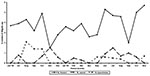
Figure. Seasonal isolates of Shigella spp. from patients with diarrhea in Indonesia (June 1998 - November 1999).
Overall, bacterial isolates of Shigella spp. were identified in 180 (5%) of 3,848 rectal swabs. The proportional contribution of S. flexneri, S. sonnei, and S. dysenteriae among shigellosis cases was 80%, 12%, and 8%, respectively. No S. boydii was detected. The percentage of representation among the three species did not vary substantially by geographic location. Notable was the reemergence of S. dysenteriae in Bali, West Kalimantan, and Batam as well as in Jakarta after a hiatus of >15 years (Table). The proportional distribution of S. flexneri, S. sonnei, and S. dysenteriae for the 5- to 12-year-old and >12-year-old groups was similar. There appeared to be no consistency in the seasonal distribution patterns of Shigella spp. (Figure). S. flexneri was the most isolated organism, followed by S. sonnei and S. dysenteriae.
Other enteric pathogens isolated were Salmonella spp. (95, 2.5%), Vibrio cholerae (80, 2.1%), V. parahaemolyticus (38, 1%), V. cholerae non-O1 (9, 0.2%), and Campylobacter spp. (27, 0.7%). Enterotoxigenic Escherichia coli were detected in 225 (18.1%) of 1,240 specimens tested by the GM1 enzyme-linked immunoassay (12). Of the 541 specimens examined for rotavirus in the age groups 0 to 5 years, 191 (35.3%) were positive. Tests to detect parasites showed Ascaris lumbricoides in 8 (2%), Blastocystis hominis in 23 (5.6%), Giardia lamblia in 3 (0.7%), and Endolimax nana in 2 (0.5%) of 407 stool specimens examined.
Clinical presentations associated with non-S. dysenteriae included abdominal cramping (79%), vomiting (56%), and fever (48%); for S. dysenteriae, percentages of the same symptoms were 100%, 64%, and 27%, respectively. Stool samples from patients with S. flexneri, S. sonnei, and S. dysenteriae were principally characterized by mucus in the absence of blood (45%) or mucus and visible blood (27%).
Overall, antibiotic susceptibility patterns showed greater resistance to ampicillin, trimethoprim-sulfamethoxazole, chloramphenicol, and tetracycline for S. flexneri (85%, 59%, 82%, and 98%, respectively, from examination of 144 isolates) and S. sonnei (32%, 79%, 37%, and 100%, respectively, from examination of 22 isolates), than for S. dysenteriae (36%, 43%, 7%, and 29%, respectively, from examination of 14 isolates). There was no evidence of resistance to ceftriaxone, norfloxacin, ciprofloxacin, or nalidixic acid, regardless of Shigella species. Antimicrobial resistance was only apparent among isolates obtained from Jakarta, Bali, and Pontianak
Our study showed that a substantial proportion (5%) of acute, debilitating diarrheal illness throughout Indonesia can be ascribed to shigellosis; moreover, S. dysenteriae was documented from various geographic locations. Both findings suggest that greater attention should be paid in highlighting the endemic and epidemic community impact of this pathogen and that laboratory detection capabilities need to be enhanced. Recognition of emerging and or reemerging disease pathogens requires reliable baseline and ongoing surveillance data.
Shifting patterns of antimicrobial-drug resistance, particularly in much of the developing world, are generally a function of overuse and misuse of antibiotic drug therapies. The spread of drug resistance is the result of poorly regulated and enforced policies. Resistance to nalidixic acid (100%) among isolates of S. dysenteriae has been reported in Bangladesh (13); however, we found no such resistance. Nevertheless, other data from Indonesia indicate increasing resistance. In a previous report (8), 72% of Shigella spp. were resistant to tetracycline, but less than 30% were resistant to chloramphenicol, trimethroprim-sulfamethoxazole, or ampicillin. Previous studies from Bangladesh and Tanzania (13,6) showed that almost all tested isolates were resistant to the antibiotics used for treatment. Similar antimicrobial resistance profiles for Shigella spp. were reported from Thailand (14), where high resistance to ampicillin, trimethroprim-sulfamethoxazole, chloramphenicol, and tetracycline was documented.
The reemergence of S. dysenteriae from several locations in Indonesia should prove cause for concern to health officials, particularly in monitoring acute, debilitating diarrheal outbreaks. The epidemic potential attributed to S. dysenteriae, as documented in Central America, Asia, and Africa, in conjunction with notably high death rates, warrants close attention to this reemerging pathogen in Indonesia (13,15,16).
Dr. Subekti is a microbiologist and medical research scientist who heads the Molecular Microbiology Section of the Enteric Diseases Program at U.S. Naval Medical Research Unit No.2, Jakarta, Indonesia.
Acknowledgment
This research was supported by the U.S. Naval Medical Research and Center Command (work unit numbers 61101A-00101.EAX 2401 and 623002A810.0101.HIX.3411).
References
- Dupont HL. Shigella. Infect Dis Clin North Am. 1998;2:599–605.
- Keusch GT, Bennish ML. Shigellosis: recent progress, persisting problems and research issues. Pediatr Infect Dis J. 1989;8:713–9. DOIPubMedGoogle Scholar
- World Health Organization. The management of bloody diarrhea in young children 1994. Geneva: World Health Organization; 1994.
- Ashkenazi S, May-Zahav M, Dinari G, Gabbay U, Zilberberg R, Samra Z. Recent trends in the epidemiology of Shigella species in Israel. Clin Infect Dis. 1993;17:897–9.PubMedGoogle Scholar
- Lee LA, Shapiro CN, Hargrett-Bean N, Tauxe RV. Hyperendemic shigellosis in the United States: a review of surveillance data for 1967-1988. J Infect Dis. 1991;64:894–900.PubMedGoogle Scholar
- Navia MM, Capitano L, Ruiz J, Vargas M, Urassa H, Schellemberg D, Typing and characterization of mechanisms of resistance of Shigella spp.isolated from feces of children under 5 years of age from Ifakara, Tanzania. J Clin Microbiol. 1999;37:3113–7.PubMedGoogle Scholar
- Simanjuntak CH, Hasibuan MA, Siregar LO, Koiman I. Microbial etiology of acute diarrhea (Indonesian) Health Study. Indonesia. 1983;10:1–9.
- Subekti D, Lesmana M, Komalarini S, Tjaniadi P, Burr D, Pazzaglia G. Enterotoxigenic Escherichia coli and other causes of infectious pediatric diarrheas in Jakarta, Indonesia. Southeast Asian J Trop Med Public Health. 1993;24:420–4.PubMedGoogle Scholar
- Simanjuntak CH, Hardjining S, Hasibuan MA, Pujarwoto Koiman I. Laboratory aspects of gastrointestinal in Indonesia, 1980-1985. Gastrointestinal Infection in Southeast SEAMIC Workshop, Japan; 1998. p. 23-31.
- Farmer JJ III, Kelly MT. Enterobacteriaceae. In: Balows A, Hausler WJ Jr, Herrmann KL, Isenberg HD, Shadomy HJ, editors. Manual of clinical microbiology. 5th ed. Washington: American Society for Microbiology; 1991. p. 360-83.
- Barry AL, Thornsberry C. Susceptibility tests: Diffusion test procedures. In: Balows A, Hausler WJ Jr, Herrmann KL, Isenberg HD, Shadomy HJ, editors. Manual of clinical microbiology. 5th ed. Washington: American Society for Microbiology; 1991. p. 1117-25.
- Svennerholm AM, Wikstrom M, Lindbland M, Holmgren J. Monoclonal antibodies against E. coli heat stable toxin (Sta) and their use in a diagnostic ST ganglioside GM1-enzyme-linked immunosorbent assay. J Clin Microbiol. 1986;24:585–90.PubMedGoogle Scholar
- Sack RB, Rahman M, Yunus M, Khan EH. Antimicrobial resistance in organisms causing diarrheal disease. Clin Infect Dis. 1997;24(Suppl 1):S102–5.PubMedGoogle Scholar
- Moolasart P. Shigellosis in Thai people. JAMA Southeast Asia. 1994;5:9–10.
- Malakooti MA, Alaii J, Shanks GD, Phillips-Howard PA. Epidemic dysentery in western Kenya. Trans R Soc Trop Med Hyg. 1997;91:541–3. DOIPubMedGoogle Scholar
- Mendizabal-Morris CA, Malta L, Gangarosa EJ, Guzman G. Epidemic Shiga-bacillus dysentery in Central America. Derivation of the epidemic and its progression in Guatemala, 1968-69. Am J Trop Med Hyg. 1971;20:927–33.PubMedGoogle Scholar
Figure
Table
Cite This ArticleTable of Contents – Volume 7, Number 1—February 2001
| EID Search Options |
|---|
|
|
|
|
|
|
Please use the form below to submit correspondence to the authors or contact them at the following address:
LCDR Buhari A Oyofo, American Embassy Jakarta, Unit 8132 NAMRU TWO, FPO, AP 965208132, Indonesia; telephone: 62 21-421-4460; fax: 62-21-424-4507LCDR Buhari A Oyofo, American Embassy Jakarta, Unit 8132 NAMRU TWO, FPO, AP 965208132, Indonesia; telephone: 62 21-421-4460; fax: 62-21-424-4507
Top