Volume 8, Number 12—December 2002
Dispatch
Isolation and Genetic Characterization of Rift Valley fever virus from Aedes vexans arabiensis, Kingdom of Saudi Arabia
Cite This Article
Citation for Media
Abstract
An outbreak of Rift Valley fever in the Kingdom of Saudi Arabia and Yemen in 2000 was the first recognized occurrence of the illness outside of Africa and Madagascar. An assessment of potential mosquito vectors in the region yielded an isolate from Aedes vexans arabiensis, most closely related to strains from Madagascar (1991) and Kenya (1997).
On September 10, 2000, accounts of unexplained hemorrhagic fever in humans and associated illness in livestock along the southwestern border of Saudi Arabia and neighboring Yemen were reported to the Ministry of Health, Kingdom of Saudi Arabia. On September 15, 2000, the Centers for Disease Control and Prevention (CDC), Atlanta, Georgia, confirmed a diagnosis of Rift Valley fever in serum samples submitted by the Ministry of Health. This confirmation marked the first occurrence of Rift Valley fever outside of Africa (1).
Rift Valley fever virus (RVFV) is an important veterinary pathogen in Africa causing abortions and deaths in young animals, primarily goats and sheep (2). This mosquito-borne virus can also infect humans by arthropod blood-feeding or by contact with infected animal fluids and tissues. RVFV infection in humans is generally not apparent or is self-limiting; serious complications including hemorrhagic fever, encephalitis, and retinitis can occur. Vector-borne virus transmission in Africa is generally associated with periodic heavy rainfall during epizootics and attendant human infections (2).
We conducted an entomologic investigation in the Asir, Jizan, and Makkah Regions, north of the suspected origin of the outbreak in Jizan, and in neighboring Yemen. Because this outbreak in the Arabian Peninsula was the first recorded outside of Africa, we wanted to determine the potential arthropod vectors and their larval habitats. A review of recent human infections indicated that the affected households were located in the foothills and at the base of the Sarawat Mountains. Four locations were selected for arthropod collections, Muhayil, Al Birk, Rijal Alma'a, and Al Majardah, because these areas are representative of the different ecologic habitats from coastal plain to mountainous regions. This fertile plain used for cultivation is known as the Tihamah.
We collected adult and immature arthropods on December 5–13, 2000, using carbon dioxide–baited CDC miniature light traps and by sampling potential larval habitats with dippers. Adult specimens were frozen in liquid nitrogen for later virus testing in Fort Collins, Colorado. To investigate potential vertical transmission of RVFV, mosquito larvae were reared to adults in the malaria control laboratory in Abha, Asir Region, for later virus testing in the United States.
All collection sites were in very arid habitats; the soil is dry and rocky, and Acacia species are the only trees present. Livestock, including goats (the predominant animal), sheep, camels, and cattle, were present at every site. In general, livestock are housed at night very close to the owners’ homes. Light traps were hung in and near the residences of recent patients, and the area was examined for larval habitats. Typical larval habitats included wastewater catchments from houses that yielded mainly Culex pipiens complex mosquitoes; pools at the edge of wadis; and small, walled, passively or actively flooded cultivated plots that yielded Aedes vexans arabiensis and Ae. vittatus (3). Ae. (Stegomyia) unilineatus, a mosquito species previously recorded from Africa, India, and Pakistan, was found in light trap collections from several sites (Godsey MS, submitted for publication). The immature and adult arthropod collections are presented in Table 1. The low species diversity and small numbers collected in light traps may have reflected that rainfall was light to nonexistent 2 weeks before our collection efforts. We did examine one walled farming plot, which contained water to a depth of approximately 5 cm; it held enormous numbers of Ae. vexans arabiensis larvae and pupae. Overnight, this habitat had dried up to a 2-m diameter pool.
Collected arthropod specimens were identified and placed into pools of up to 50 individual mosquitoes by collection site. A total of 161 pools were triturated, clarified by centrifugation, and spread onto confluent sheets of Vero cells in six-well plates (4). A single pool (SA01 #1322) yielded a virus isolate. The virus was identified as RVFV by sequencing a DNA fragment amplified from the M segment by reverse transcription-polymerase chain reaction using the primers RVF3082 5′actttgtgggagcagccgtatctt3′ and RVF3400 5′cctgcttcccgcctatcatcaaat3′.
RVFV was isolated from a pool of 37 Ae. vexans arabiensis female mosquitoes collected by light trap at site 3 (N18° 45.089 min; E41° 56.373 min) near the city of Muhayil. A human infection was recorded from this site ("Agida"); we also witnessed aborted bovine fetuses on the property. Other arthropods collected at this site included Cx. tritaeniorhynchus and psychodid sandflies.
Figure
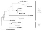
RVFV is a member of the virus family, Bunyaviridae; the genomes of these viruses exist in three pieces or segments: small (S), medium (M), and large (L). We sequenced a portion of each of the genome segments and analyzed them by the maximum likelihood algorithm in PAUP (5) in relation to the published sequences of other geographic isolates to determine the possible origin of the mosquito isolate and whether the isolate was a possible reassortant between two existing virus strains (5,6). Maximum likelihood trees for each genomic segment shared identical topologies (data not shown). The congruence of placement of the Saudi virus strain in the three trees indicated that this virus was not a reassortant. The phylogram for segment M (Figure; Table 2) demonstrates that the most closely related RVFV isolates were from Kenya (1997) and Madagascar (1991). Reasonable hypotheses to explain how RVFV was introduced into the Kingdom of Saudi Arabia (and/or Yemen) from East Africa are that an infected mosquito was carried over the narrow waterway between the Red Sea and the Gulf of Aden by air currents or that infected livestock were imported from East Africa. How long the virus was in the Arabian Peninsula before the epidemic occurred is unknown.
The most abundant culicine mosquitoes we collected were Ae. vexans arabiensis, Cx. pipiens complex, and Cx. tritaeniorhynchus. All three species should be considered important epidemic and epizootic vectors of RVFV in Saudi Arabia. The floodwater mosquito, Ae. vexans arabiensis, has the potential to be an important epidemic and epizootic vector because of the tremendous numbers of individual mosquitoes that are produced after a flooding rain. Whether or not RVFV is able to persist on the Arabian Peninsula is unknown. Clearly, vertical transmission of the virus in the epidemic mosquito vector would be an important factor to consider.
Dr. Miller is chief of the Virus and Vector Molecular Biology Section, Arbovirus Diseases Branch, Division of Vector-Borne Infectious Diseases, Centers for Disease Control and Prevention. His research interests include arboviral ecology, virus-vector interactions, and molecular systematics.
Acknowledgment
We thank Abdullah I. Al-Sharif, Abdullah Abu-Dahish, A.M. Khan, and Mr. Maray Yahya for their generous logistical assistance. We would also like to thank Amy Lambert for doing the original reverse transcription-polymerase chain reaction on the isolate.
References
- Centers for Disease Control and Prevention. Outbreak of Rift Valley fever—Saudi Arabia, August–October, 2000. MMWR Morb Mortal Wkly Rep. 2000;49:905–8.PubMedGoogle Scholar
- Peters CJ. Emergence of Rift Valley fever. In: Saluzzo JF, Dodet B, editors. Factors in the emergence of arboviruses. Paris: Elsevier; 1997. p. 253–64.
- White GB. Notes on a catalog of Culicidae of the Ethiopian region. Mosquito Systematics. 1975;7:303–44.
- Miller BR, Nasci RS, Lutwama JJ, Godsey MS, Savage HM, Lanciotti RS, First field evidence for natural vertical transmission of West Nile virus in Culex univittatus complex mosquitoes from Rift Valley Province, Kenya. Am J Trop Med Hyg. 2000;62:240–6.PubMedGoogle Scholar
- Swofford DL. PAUP*:Phylogenetic Analysis Using Parsimony (*and other methods). Version 4.08b. Sunderland (MA): Sinauer Associates; 1998.
- Sall AA, de A. Aanotto PM, Sene OK, Zeller HG, Digoutte JP, Thiongane Y, . Genetic reassortment of Rift Valley fever in nature. J Virol. 1999;73:8196–200.PubMedGoogle Scholar
Figure
Tables
Cite This ArticleTable of Contents – Volume 8, Number 12—December 2002
| EID Search Options |
|---|
|
|
|
|
|
|
Please use the form below to submit correspondence to the authors or contact them at the following address:
Barry R. Miller, Division of Vector-Borne Infectious Diseases, Centers for Disease Control and Prevention, P.O. Box 2087, Fort Collins, CO 80522, USA; fax: 970-221-6476;
Top