Volume 8, Number 4—April 2002
Research
Biofilm on Ventriculo-Peritoneal Shunt Tubing as a Cause of Treatment Failure in Coccidioidal Meningitis
Figure 2
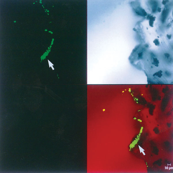
Figure 2. A. Scanning electron microscopy shows the presence of leukocytes and red blood cells on the tip of the ventriculo-peritoneal mass, within which coccoid cells can be visualized. The enclosing matrix material has condensed by dehydration, but the outline of the 4- to 6-μm coccoid cells (arrow), similar to those of C. immitis, can be resolved within the mass (x4,000). B. Scanning electron microscopy of the surface of the ventriculo-peritoneal shunt, showing complete colonization of the surface by a matrix-enclosed biofilm formed by the cells of C. immitis. Within the dehydration-condensed matrix of this biofilm, a hyphal element (arrow) and coccoid cells (4-6 μm) of the pathogen can be discerned (x5,000).
Page created: July 15, 2010
Page updated: July 15, 2010
Page reviewed: July 15, 2010
The conclusions, findings, and opinions expressed by authors contributing to this journal do not necessarily reflect the official position of the U.S. Department of Health and Human Services, the Public Health Service, the Centers for Disease Control and Prevention, or the authors' affiliated institutions. Use of trade names is for identification only and does not imply endorsement by any of the groups named above.