Skip directly to site content Skip directly to page options Skip directly to A-Z link Skip directly to A-Z link Skip directly to A-Z link
Volume 14, Number 6—June 2008
Letter

Splenic Rupture and Malignant Mediterranean Spotted Fever

Laura Schmulewitz*, Kaoutar Moumile*, Natacha Patey-Mariaud de Serre*, Sylvain Poirée*, Edith Gouin†, Frédéric Mechaï*, Véronique Cocard*, Marie-France Mamzer-Bruneel*, Eric Abachin*, Patrick Berche*, Olivier Lortholary*†, and Marc Lecuit*†Comments to Author 
Author affiliations: *Université Paris Descartes, Hôpital Necker-Enfants Malades, Paris, France; †Institut Pasteur, Paris, France;

Main Article

Figure

Histopathologic and immunohistochemical labelings of spleen and skin tissue samples. Tissue samples were fixed in 10% formalin, paraffin-embedded, and examined after hematoxylin-eosin staining, Gimenez staining, or immunostaining with the R47 anti-Rickettsia conorii polyclonal rabbit antibody. The spleen red pulp indicated congestion and ill-defined nodules varying in size and comprising macrophages, polymorphonuclear neutrophils, and necrotic cells (A, magnification ×100). A diffuse macrophage

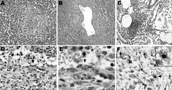
Figure. Histopathologic and immunohistochemical labelings of spleen and skin tissue samples. Tissue samples were fixed in 10% formalin, paraffin-embedded, and examined after hematoxylin-eosin staining, Gimenez staining, or immunostaining with the R47 anti-Rickettsia conorii polyclonal rabbit antibody. The spleen red pulp indicated congestion and ill-defined nodules varying in size and comprising macrophages, polymorphonuclear neutrophils, and necrotic cells (A, magnification ×100). A diffuse macrophage infiltration with abundant hemophagocytosis (not shown) and venulitis (B, magnification ×50) was also observed. In the skin, leukocytoclastic vasculitis with focal vascular necrosis and nonocclusive luminal thrombi were noted in dermal capillaries (C, magnification ×100). Intracellular images evocative of rickettsiae were observed in the splenic arteriolar endothelium upon immunohistochemical staining (D, arrow, magnification ×200; magnified view shown in E, arrowhead, magnification ×500). No infected cells were observed in nodular inflammatory splenic lesions. Immunohistochemical staining also disclosed intracellular immunolabeled dots in cells that could correspond to infected dermal macrophages (F, arrowhead, magnification ×300; magnified view shown in inset, magnification ×600), at a distance from the vascular alterations. Endothelial cells of dermal capillaries were also immunolabeled (Appendix Figure).

Main Article

Page created: July 09, 2010
Page updated: July 09, 2010
Page reviewed: July 09, 2010
The conclusions, findings, and opinions expressed by authors contributing to this journal do not necessarily reflect the official position of the U.S. Department of Health and Human Services, the Public Health Service, the Centers for Disease Control and Prevention, or the authors' affiliated institutions. Use of trade names is for identification only and does not imply endorsement by any of the groups named above.
file_external