Volume 14, Number 9—September 2008
Dispatch
Neurobrucellosis in Stranded Dolphins, Costa Rica
Gabriela Hernández-Mora, Rocío González-Barrientos, Juan-Alberto Morales, Esteban Chaves-Olarte, Caterina Guzmán-Verri, Elías Baquero-Calvo, María-Jesús De-Miguel, Clara-María Marín, José-María Blasco, and Edgardo Moreno
Figure 1
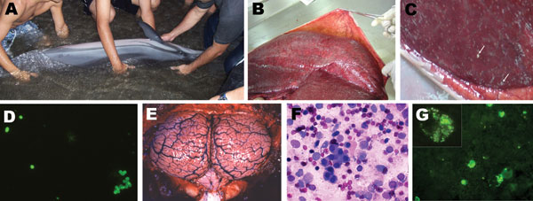
Figure 1. Clinical, pathologic, and immunofluorescence findings in stranded striped dolphin, Stenella coeruleoalba. A) Striped dolphin displaying swimming disorders being assisted by local persons; B) dolphin fetus within placenta; C) punctuated placental abscesses (arrows); D) immunofluorescent brucellae in impressions of placenta tissues; E) congested and hyperemic brain and cerebellum; F) Wright-Giemsa–stained mononuclear cell infiltrate in cerebrospinal fluid; G) immunofluorescent green Brucella spp. and Brucella debris within phagocytic cells infiltrating cerebrospinal fluid; the inset corresponds to an amplified phagocytic cell with fluorescent Brucella spp. and debris.
Page created: July 13, 2010
Page updated: July 13, 2010
Page reviewed: July 13, 2010
The conclusions, findings, and opinions expressed by authors contributing to this journal do not necessarily reflect the official position of the U.S. Department of Health and Human Services, the Public Health Service, the Centers for Disease Control and Prevention, or the authors' affiliated institutions. Use of trade names is for identification only and does not imply endorsement by any of the groups named above.