Volume 17, Number 12—December 2011
Letter
Iridovirus Infection in Chinese Giant Salamanders, China, 2010
Wuzi Dong, Xiaoming Zhang, Changming Yang, Junhui An, Jinzhou Qin, Fengfeng Song, and Wenxian Zeng
Figure
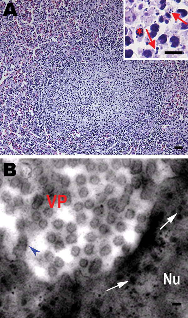
Figure. Histologic changes in the spleen of sick Chinese giant salamanders (Andrias davidianus), People’s Republic of China, 2010. Electron microscopy shows virus particles in splenocytes. A) Hyperplasia of lymphoid nodules in the splenic white pulp. Inset: Some splenocytes contain nuclear debris (arrows) and macrophages (asterisk). Hematoxylin and eosin stain; scale bars = 80 μm. B) Electron microscopy image of viral particles in splenocytes. Many viral particles are cytoplasmic and appear hexagonal or round. Scale bar = 200 nm. VP, viral particles in cytoplasm; Nu, nucleus; arrowhead, provirus in nuclear membrane; arrows, provirus in nucleus.
Page created: November 30, 2011
Page updated: November 30, 2011
Page reviewed: November 30, 2011
The conclusions, findings, and opinions expressed by authors contributing to this journal do not necessarily reflect the official position of the U.S. Department of Health and Human Services, the Public Health Service, the Centers for Disease Control and Prevention, or the authors' affiliated institutions. Use of trade names is for identification only and does not imply endorsement by any of the groups named above.