Volume 18, Number 11—November 2012
Research
Mycoplasmosis in Ferrets
Matti Kiupel , Danielle R. Desjardins, Ailam Lim, Carole Bolin, Cathy A. Johnson-Delaney, James H. Resau, Michael M. Garner, and Steven R. Bolin
Figure 5
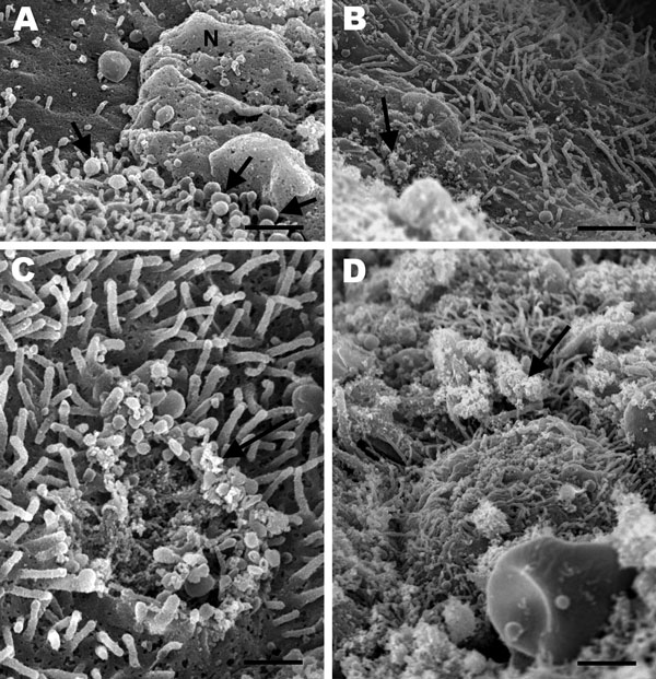
Figure 5. . . Scanning electron micrographs of the lung from a 2-year-old ferret that died of acute dyspnea, showing A) marked loss of cilia with multifocal degenerative changes characterized by bulbous swelling of cilia (arrows) and necrosis of bronchial epithelial cells (N) (scale bar = 1 µm); B) marked loss of cilia and numerous pleomorphic mycoplasma-like organisms diffusely attached to the mucosal surface (arrow) (scale bar = 1.25 µm); C) focal area of cilia loss and cell membrane damage with mycoplasma-like organisms (arrow) at the periphery of the lesion (scale bar = 400 nm); and D) many mycoplasma-like organisms (arrow) covering ciliated bronchial epithelial cells (scale bar = 2 µm).
Page created: October 15, 2012
Page updated: October 15, 2012
Page reviewed: October 15, 2012
The conclusions, findings, and opinions expressed by authors contributing to this journal do not necessarily reflect the official position of the U.S. Department of Health and Human Services, the Public Health Service, the Centers for Disease Control and Prevention, or the authors' affiliated institutions. Use of trade names is for identification only and does not imply endorsement by any of the groups named above.