Skip directly to site content Skip directly to page options Skip directly to A-Z link Skip directly to A-Z link Skip directly to A-Z link

Disclaimer: Early release articles are not considered as final versions. Any changes will be reflected in the online version in the month the article is officially released.

Volume 32, Number 7—July 2026

Research Letter

New World Ocular Dirofilariasis Caused by Dirofilaria repens Infection, United States

Ben J. GlasgowComments to Author , Mackenzie Collins, Luke Helminiak, Joshua A. Lieberman, Blaine A. Mathison, and Shangxin Yang
Author affiliation: University of California Los Angeles David Geffen School of Medicine, Los Angeles, California, USA (B.J. Glasgow, M. Collins, L. Helminiak, S. Yang); University of Washington, Seattle, Washington, USA (J.A. Lieberman); ARUP Laboratories and University of Utah, Salt Lake City, Utah, USA (B.A. Mathison)

Main Article

Figure 1

Images from patient with ocular dirofilariasis caused by Dirofilaria repens in California, USA. A) Axial plane of T-2–weighted orbital magnetic resonance imaging revealing a well circumscribed cystic lesion in the left lower eyelid. The center of the lesion is hyperintense and circumscribed by a hypointense signal. The interpretation was a benign inflammatory lesion. B) Histopathologic image from hematoxylin and eosin–stained slide that reveals a 500-micron diameter cross section of a nematode with a thick cuticular wall featuring external cuticular ridges (indicated by r) and vertically oriented muscle (indicated by m) extending toward the internal cavity (coelomyarian) that are numerous per quadrant (polymyarian), as well as paired reproductive tubes (indicated by t), and simple intestine (indicated by i).

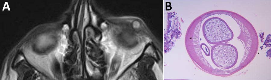
Figure 1. Images from patient with ocular dirofilariasis caused by Dirofilaria repens in California, USA. A) Axial plane of T-2–weighted orbital magnetic resonance imaging revealing a well circumscribed cystic lesion in the left lower eyelid. The center of the lesion is hyperintense and circumscribed by a hypointense signal. The interpretation was a benign inflammatory lesion. B) Histopathologic image from hematoxylin and eosin–stained slide that reveals a 500-micron diameter cross section of a nematode with a thick cuticular wall featuring external cuticular ridges (indicated by r) and vertically oriented muscle (indicated by m) extending toward the internal cavity (coelomyarian) that are numerous per quadrant (polymyarian), as well as paired reproductive tubes (indicated by t), and simple intestine (indicated by i).

Main Article

Page created: May 28, 2026
Page updated: June 23, 2026
Page reviewed: June 23, 2026
The conclusions, findings, and opinions expressed by authors contributing to this journal do not necessarily reflect the official position of the U.S. Department of Health and Human Services, the Public Health Service, the Centers for Disease Control and Prevention, or the authors' affiliated institutions. Use of trade names is for identification only and does not imply endorsement by any of the groups named above.
file_external