Volume 10, Number 5—May 2004
Dispatch
Mycobacterium africanum Cases, California
Cite This Article
Citation for Media
Abstract
Five Mycobacterium tuberculosis complex isolates in California were identified as M. africanum by spoligotyping, single nucleotide polymorphisms, a deletion mutation, and phenotypic traits, confirming it as a cause of tuberculosis in the United States. Three of the five patients from whom M. africanum was isolated had lived in Africa.
Mycobacterium africanum is a member of the M. tuberculosis complex, which has been isolated from humans in equatorial Africa. The disease produced by M. africanum is similar to that caused by M. tuberculosis or M. bovis, and like M. tuberculosis, this organism is likely spread by aerosol transmission (1). Human tuberculosis caused by M. africanum has been reported in Europe (2,3). However, we are unaware of previous reports of disease caused by M. africanum in the United States.
M. africanum may be identified by spoligotyping (4), by specific deletion mutations (5), DNA fingerprinting by IS6110 restriction fragment length polymorphisms (RFLP) (4), or a combination of these methods. Isolates were initially identified as M. tuberculosis complex by using the AccuProbe system (Gen-Probe; San Diego, CA). The isolates then underwent IS6110-based RFLP fingerprinting. The RFLP analyses were performed according to the method of van Embden et al. (6). In addition to providing genotyping results, RFLP fingerprinting confirmed the identification obtained with Accuprobe.
All strain typing was performed in-house at the Microbial Diseases Laboratory, California Department of Health Services. This laboratory has compiled a database (Genomic Solutions BioImage) of approximately 7,000 DNA fingerprints, typed by IS6110 RFLP (6,7) from throughout California; most are from the San Francisco Bay area. Three isolates (from patients A, B, and C) were initially suspected of being M. africanum because of an epidemiologic association with Africa. These isolates were fingerprinted by IS6110 RFLP and by spoligotyping (8). All three were found to have the “signature” spoligotype described by Viana-Niero et al. as being characteristic of M. africanum (4), namely, they were missing spacers 8, 9, and 39 but had spacers 40–43. Using BioImage software, we searched the laboratory’s database for IS6110 fingerprints that matched those of the three cases with an African connection. This search yielded an additional two matches, cases D and E. Isolates from cases D and E were then genotyped by using spoligotyping and found to have the M. africanum signature spoligotype.
The five M. africanum isolates were further characterized by performing standard biochemical identification tests and testing for susceptibility to pyrazinamide (PZA). Niacin production and nitrate reduction were detected as described by Kent and Kubica (9). Susceptibility to PZA was determined by using the BACTEC radiometric assay performed according to the method of Salfinger et al. (10). The M. africanum isolates were then examined to determine whether they had the RD9 deletion and specific oxyR and katG sequence mutations.
Brosch et al. had reported that isolates of M. tuberculosis do not have the RD9 deletion, whereas other members of the M. tuberculosis complex, including M. bovis, M. microti, and M. africanum have this deletion (5). Two sets of primers for detecting the RD9 deletion, designed as described by Brosch et al., were obtained from Alex Pym at Stanford University. A ≈480-bp region was amplified by polymerase chain reaction (PCR) from M. africanum by using the flanking primers, and a ≈375-bp region was amplified from M. tuberculosis by using the internal primers. After an initial denaturation step of 10 min at 95°C, amplification was performed for 35 cycles at 95°C for 30 s, 55°C for 1 min, and 72°C for 4 min, and a final step of elongation at 72°C for 10 min. The final concentration of each component in the 50-μL PCR reaction tube was 1x PCR buffer, 2.5 mmol/L of MgCl2, and 1.5 U of AmpliTaq Gold polymerase using AmpliTaq Gold kit with GeneAmp 10X PCR buffer II and MgCl2 solution (Applied Biosystems, Foster City, CA), 1.25 mmol/L of deoxynucleotide triphosphate mix (Applied Biosystems), 0.2 μmol/L of each primer, 5 μL of DNA template. A M. tuberculosis strain, H37Rv, and M. africanum strain ATCC 25420 were included in PCR runs as reference strains.
A 548-bp region of the oxyR locus was amplified by PCR using the primer sequences described by Sreevatsen et al. (11). After an initial denaturation step of 4 min at 95°C, amplification was performed for 30 cycles with the following parameters: 95°C for 30 s, 55°C for 30 s, and 72°C for 45 s. A final elongation step was performed for 5 min at 72°C. For katG, a primer set, 5′-TGCTGGCGCTTGGCAATACA and 5′-GCCGCGCTTGTCGCTACC, was designed to amplify a 429-bp region encompassing codon 463. With the exception of an annealing temperature of 60°C, the amplification parameters for katG were identical to those described for oxyR. The amplified products were purified by using QIAquick spin columns (Qiagen Inc., Valencia, CA) and subjected to dRhodamine Terminator Cycle Sequencing (Applied Biosystems, Foster City, CA) as recommended by the manufacturer. The cycle sequencing reactions were analyzed on an ABI 377 DNA sequencer, and the sequences aligned with those derived from M. tuberculosis strain H37Rv and M. africanum strain ATCC 25420.
Figure
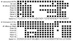
Figure. Spacer oligonucleotide typing (spoligotyping) results for cultures from patients A through E, together with representative patterns from stock cultures of Mycobacterium tuberculosis complex species. Spoligotypes for patients C and E are...
For case histories, see Table 1. The five isolates were identified as M. tuberculosis complex by a positive result with the Gen-Probe AccuProbe system and by the presence of IS6110 insertion sequences. When spoligotyping was performed on isolates from cases A through E, all were found to have the M. africanum signature spoligotype, as described by Viana-Niero et al. (4). The results of spoligotyping are shown in the Figure.
The five isolates were M. africanum, based on several criteria shown in Table 2. Susceptibility of the strains to PZA, nitrate reduction, production of niacin, and the presence of guanosine at oxyR285 were incompatible with identification of the isolates as M. bovis. The RD9 deletion was shown for all five isolates and the M. africanum reference strain, ATCC 25420, by 1) the presence of a ≈480-bp band on the agarose gel when the PCR was performed with the flanking primers and 2) the absence of a ≈375-bp band when the PCR was performed with the internal primers. The RD9 deletion was incompatible with identification of the isolates as M. tuberculosis, and CTG at katG463 suggested that the isolates were not M. tuberculosis, since most isolates of this species have CGG at this site (7).
David et al. (12) and Frothingham et al. (13) reported that some isolates of M. africanum are resistant to PZA and fail to accumulate niacin, distinguishing them from most strains of M. tuberculosis, while other isolates are susceptible to PZA and produce niacin. These five isolates of M. africanum were susceptible to PZA, and three out of the five tested positive for niacin production and nitrate reduction (the remaining two cultures had lost viability and were not available for this testing). This rendered the cultures phenotypically indistinguishable from M. tuberculosis (12–14). For each of the reported cases, the signature M. africanum spoligotype pattern described by Viana-Niero et al. (4) was supported by the single nucleotide polymorphisms characteristic of M. africanum and by the RD9 mutation that characterizes members of the tuberculosis complex other than M. tuberculosis.
At present, the Microbial Diseases Laboratory plans to spoligotype all strains of M. tuberculosis complex. Clinical features and drug susceptibility were not distinctive for M. africanum isolates, nor were all associated with patients who came from Africa. No connection with a source case from Africa was found for patients D or E. However, since spoligotyping will be routine, noting the signature africanum spoligotype may be worthwhile. This spoligotype may indicate an increased chance that the patient may have acquired the M. tuberculosis complex infection in Africa or possibly from an African source case.
Dr. Desmond has worked at the Microbial Diseases Laboratory, California Department of Health Services, since 1990. His research interests include laboratory methods for detecting drug resistance in Mycobacterium tuberculosis and applications for molecular strain typing in the mycobacteriology laboratory.
References
- Adler JJ, Rose DN. Transmission and pathogenesis of tuberculosis. In: Rom WN, Garay SM, editors. Tuberculosis. Boston: Little, Brown, and Co.; 1996. p. 129–40.
- Grange JM, Yates MD. Incidence and nature of human tuberculosis due to Mycobacterium africanum in southeast England. Epidemiol Infect. 1989;103:127–32. DOIPubMedGoogle Scholar
- Grosset J, Decroix G, Sors C. Tuberculosis due to Mycobacterium africanum in African Negroes in the Paris area. Rev Tuberc Pneumol (Paris). 1971;35:430–6.PubMedGoogle Scholar
- Viana-Niero C, Gutierrez C, Sola C, Filliol I, Boulahbal F, Vincent V, Genetic diversity of Mycobacterium africanum clinical isolates based on IS6110-restriction fragment length polymorphism analysis, spoligotyping, and variable number of tandem repeats. J Clin Microbiol. 2001;39:57–65. DOIPubMedGoogle Scholar
- Brosch R, Gordon SV, Marmiesse M, Brodin P, Buchrieser C, Eiglmeier K, A new evolutionary scenario for the Mycobacterium tuberculosis complex. Proc Natl Acad Sci U S A. 2002;99:3684–9. DOIPubMedGoogle Scholar
- van Embden JDA, Cave MD, Crawford JT, Dale JW, Eisenach KD, Gicquel B, Strain identification of Mycobacterium tuberculosis by DNA fingerprinting: recommendations for a standardized methodology. J Clin Microbiol. 1993;31:406–9.PubMedGoogle Scholar
- Mazurek GH, Cave MD, Eisenach KD, Wallace RJ, Bates JH, Crawford JT. Chromosomal DNA fingerprint patterns produced with IS6110 as strain-specific markers for epidemiologic study of tuberculosis. J Clin Microbiol. 1991;29:2030–3.PubMedGoogle Scholar
- Kamerbeek J, Schouls L, Kolk A, van Agterveld M, van Soolingen D, Kuijper S, Simultaneous detection and strain differentiation of Mycobacterium tuberculosis for diagnosis and epidemiology. J Clin Microbiol. 1997;35:907–14.PubMedGoogle Scholar
- Kent P, Kubica G. Public health mycobacteriology: a guide for the level III laboratory. Atlanta: Centers for Disease Control; 1985.
- Salfinger M, Reller LB, Demchuk B, Johnson ZT. Rapid radiometric method for pyrazinamide susceptibility testing of Mycobacterium tuberculosis. Res Microbiol. 1989;140:301–9. DOIPubMedGoogle Scholar
- Sreevatsen S, Escalante P, Pan X, Gillies DA II, Siddiqui S, Khalaf CN, Identification of a polymorphic nucleotide in oxyR specific for Mycobacterium bovis. J Clin Microbiol. 1996;34:2007–10.PubMedGoogle Scholar
- David HL, Jahan M-T, Jumin A, Grandry J, Lehman EH. Numerical taxonomy analysis of Mycobacterium africanum. Int J Syst Bacteriol. 1978;28:467–72. DOIGoogle Scholar
- Frothingham R, Strickland PL, Bretzel G, Ramaswamy S, Musser JM, Williams DL. Phenotypic and genotypic characterization of Mycobacterium africanum isolates from West Africa. J Clin Microbiol. 1999;37:1921–6.PubMedGoogle Scholar
- Niemann S, Richter E, Rusch-Gerdes S. Differentiation among members of the Mycobacterium tuberculosis complex by molecular and biochemical features: evidence for two pyrazinamide-susceptible subtypes of M. bovis. J Clin Microbiol. 2000;38:152–7.PubMedGoogle Scholar
Figure
Tables
Cite This ArticleTable of Contents – Volume 10, Number 5—May 2004
| EID Search Options |
|---|
|
|
|
|
|
|
Please use the form below to submit correspondence to the authors or contact them at the following address:
Edward Desmond, Microbial Diseases Laboratory, California Department of Health Services, 850 Marina Bay Parkway, Richmond, CA 93804, USA; fax: 510-412-3706
Top