Volume 11, Number 6—June 2005
Dispatch
Dana Cole, Georgia Division of Public Health, Notifiable Disease Section, Department of Human Resources, 2 Peachtree Free-living Canada Geese and Antimicrobial Resistance
Cite This Article
Citation for Media
Abstract
We describe antimicrobial resistance among Escherichia coli isolated from free-living Canada Geese in Georgia and North Carolina (USA). Resistance patterns are compared to those reported by the National Antimicrobial Resistance Monitoring System. Canada Geese may be vectors of antimicrobial resistance and resistance genes in agricultural environments.
The epidemiology of zoonotic diseases is growing in scope and importance in public health as the interface between animal and human habitats narrows and new diseases emerge. Historically, zoonotic disease research has emphasized occupational or animal-origin foodborne exposures. However, environmental exposure pathways to zoonotic pathogens are increasingly documented as foodborne disease surveillance and control efforts prove successful. Nonanimal-origin sources of zoonotic infection, such as raw fruits and vegetables, nuts, and water, are reported more often (1–3). Although fecal contamination of raw food products in fields is an important source of zoonotic infection (1), the source of contamination is usually not determined. Consequently, environmental reservoirs of microbes of public health importance need to be investigated.
Figure
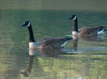
Figure. . Free-living populations of Canada Geese (Branta canadensis) can serve as reservoirs of antimicrobial-resistant bacteria such as Escherichia coli..
Canada Geese (Branta canadensis) (Figure) populations have steadily increased in the past 50 years and have become a nuisance in some areas (4,5). The large amount of feces produced by geese congregating around surface water bodies is a source of environmental contamination and, potentially, zoonotic pathogens (4–7). Feces from large flocks are major contributors to fecal coliform levels in reservoirs that supply drinking water for some cities (5,6), and free-living bird populations can serve as reservoirs for pathogenic bacteria such as Salmonella (8,9), Escherichia coli (10), Campylobacter (10,11), Listeria (11), and Chlamydia (9). Thus, wild bird populations can amplify and eventually transmit infectious microbes to humans by directly contaminating agricultural fields or surface waters used for drinking, recreation, or crop irrigation.
Free-living and domestic bird populations can also be reservoirs of drug-resistant bacterial pathogens or resistant genetic elements. Antimicrobial-resistant organisms in domestic animals such as poultry, beef, and swine are well documented (12,13) and have been implicated as reservoirs for multidrug-resistant foodborne pathogens. Interaction with waste materials from these livestock species may confer resistant pathogens and genetic elements to free-ranging wildlife, potentially creating an additional environmental reservoir of resistant organisms (8). We examine the impact of habitat on antimicrobial susceptibilities of E. coli isolates recovered from different flocks of resident, free-living Canada Geese to determine the potential for these animals to be additional sources of antimicrobial resistance through exposure pathways that originate in the environment.
Collaborators from separate regions collected cloacal swabs or fresh guano from Canada Geese at 4 geographically diverse surface water bodies in Georgia (n = 72) and North Carolina (n = 90). Cloacal swabs were taken from each of 24 Canada Geese captured at each Georgia site. At the North Carolina site, groups of resident geese were followed by a study investigator on 8 occasions, and fresh guano was collected from 7 to 8 birds. Geographic locations represented the following land uses: recreational (Stone Mountain Park, GA), agricultural (Craven County, NC; Griffin, GA), and industrial (Lake Juliette, GA). The Craven County site was near a swine housing facility, and geese were observed using swine waste lagoons and adjacent surface waters and farm fields. The Griffin site was not near food animal facilities but was adjacent to test plots used by crops and soils scientists at the University of Georgia Experiment Station. Neither the Griffin Lake nor the lake at Stone Mountain Park is downstream from a sewage treatment plant; however, Lake Juliette, a reservoir for an electrical generation plant, is formed by Rum Creek and water pumped from the Ocmulgee River, on which several sewage treatment facilities reside.
Isolation and biochemical identification of E. coli from free-ranging geese were performed as follows. In Georgia, cloacal swabs were transported to the investigators' laboratory and stored at 4°C. The following day, swabs were used to inoculate brain heart infusion broth (BHIB) and incubated overnight at 37°C. BHIB cultures were subsequently streaked for isolated colonies on MacConkey agar plates; 1 lactose-fermenting colony was selected from each goose sample that exhibited growth on agar. In North Carolina, bird samples were pooled and transported to the laboratory on ice in sterile, plastic bottles and stored at 4°C overnight. A 1-g sample of guano was diluted in sterile phosphate-buffered saline, and 3 separate dilutions were filtered through 47-mm, 0.45-μm pore-size, gridded cellulose ester filters. After overnight culture on mFC agar plates at 37°C, filters containing a countable number of fecal coliform colonies (20–100 CFUs) were transferred to plates containing nutrient agar and 4-methylumbelliferyl-β-D-glucuronide (MUG) and incubated for an additional 3–4 h. Colonies fluorescing blue under long wavelength UV light were selected for biochemical confirmation. Up to 5 presumptive E. coli colonies were selected from each sampling round. All isolates were identified to genus and species level with the Vitek System (bioMériux Vitek, Hazelwood, MO, USA).
Antimicrobial-susceptibility patterns for each confirmed bacterial isolate were determined by broth microdilution with the Sensititre automated antimicrobial susceptibility system (Trek Diagnostic Systems, Westlake, OH, USA) and interpreted according to NCCLS criteria for dilution susceptibility testing methods when applicable (14). Antimicrobial susceptibilities were assessed for amikacin, amoxicillin/clavulanic acid, ampicillin, apramycin, cefoxitin, ceftiofur, ceftriaxone, cephalothin, chloramphenicol, ciprofloxacin, gentamicin, imipenem, kanamycin, nalidixic acid, streptomycin, sulfamethoxazole, tetracycline, and trimethoprim/sulfamethoxazole. E. coli ATCC 25922, E. coli ATCC 35218, and Pseudomonas aeruginosa ATCC 27853 were quality-control organisms. Differences in the proportion of resistant isolates were analyzed by chi-square test (SAS, ver. 8.01, SAS Institute Inc., Cary, NC, USA).
Isolates were also screened for class 1 integrase gene intI1 and integron-associated antimicrobial resistance genes sul1 and aadA1 by polymerase chain reaction (PCR). Isolates that exhibited resistance to β-lactam antimicrobial agents were screened by PCR for TEM β-lactamase gene, blaTEM, with appropriate positive and negative control strains. DNA template for PCR was prepared as described by Hilton et al. (15).
The Table describes antimicrobial resistance phenotypes and associated resistance determinants in E. coli isolates recovered from Canada Geese stratified by geographic site. No gram-negative enteric bacteria were isolated from geese at Stone Mountain Park, although E. coli isolates were recovered from the bird waste from Griffin, Lake Juliette, and Craven County. The number of isolates recovered was much higher among geese in agricultural areas compared to other land usages (e.g., industrial or recreational). The proportion of isolates resistant to antimicrobial agents was significantly greater (p = 0.0004) among E. coli isolates from Craven County geese, where interaction with swine waste lagoons was observed. Antimicrobial resistance patterns in this population matched those most commonly reported for swine Enterobacteriaceae from the National Antimicrobial Resistance Monitoring System (NARMS) studies (e.g., tetracycline, streptomycin, sulfamethoxazole, and ampicillin resistance) (12,13). Most E. coli isolates (72%) recovered from Craven County geese exhibited resistance to ≥1 antimicrobial agent. In contrast, resistant E. coli recovered from agricultural geese in Georgia (Griffin) with no apparent contact with livestock wastes had a lower proportion of resistance (19%) and only exhibited resistance to β-lactam antimicrobial agents (cefoxitin-amoxicillin/clavulanic acid-cephalothin).
All E. coli isolates, except those from Craven County, were negative for class 1 integrons. Forty-four percent of E. coli isolates (n = 25) from Craven County Canada Geese possessed ≥1 antimicrobial-resistant determinant. Nine E. coli isolates were positive for class 1 integrase gene intI1; 6 isolates possessed a TEM β-lactamase.
Outbreaks of illness associated with raw food products have been increasing, in part because of increased human consumption of fresh produce (1). However, several sources of preharvest contamination have been identified, including fecal material, contaminated irrigation water, and wild fowl (1). In a previous study, 32% of the Salmonella isolates from wild birds submitted to the Southeastern Cooperative Wildlife Disease Study were resistant to sulfamethoxazole, and 18.1% were resistant to both sulfamethoxazole and streptomycin (8). These findings are likely a result of interaction of these populations with environmental sources of enteric bacteria. In our study, the spectrum of E. coli resistance was very different among agricultural habitat geese, depending upon their exposure to livestock wastes. With growing populations of Canada Geese and associated evidence that they contribute to microbial water contamination (5,6), we hypothesized that observed resistance patterns might be related to the anthropogenic land usage of the bird habitats and that Canada Geese could serve as a vector of antimicrobial resistance genes between sources of fecal wastes and other environmental media. Little or no resistance was observed among the E. coli isolates recovered from Canada Geese in regions with no known direct contact with liquid wastes. However, geese in direct contact with liquid swine wastes had a significantly higher prevalence of antimicrobial resistance. Comparing these data with those reported recently by NARMS shows similar resistance profiles between E. coli isolates recovered from Canada Geese in contact with livestock wastes (Craven County) and those recovered from both food animals and fresh fruits and vegetables (12,13). In addition, a substantial number of isolates from several Canada Geese that had direct contact with lagoons containing liquid swine waste carried integrons and their associated resistance genes.
This and other studies suggest that resident, free-living, and migratory birds can be potential vectors of zoonotic pathogens, including antimicrobial-resistant variants, between waste-handling facilities and other agricultural resources, such as crops and water. Although all of our study populations of Canada Geese were nonmigratory, this species could serve to disperse bacteria between widely separated locations. In addition, since these birds use farm ponds and waste lagoons and graze on pastures inhabited by cattle and other livestock, the opportunities exist for new health problems in wildlife populations to emerge as well as new reservoirs of zoonotic disease to form. This work is the basis of continuing efforts to examine the potential role of wildlife in agricultural habitats as vectors of antimicrobial resistance in the environment.
This work was supported by USDA NRICGP grants 99-35212-8680 and 01-35212-10877 (Georgia) and USDA NRI Grant 99-35102-8178, USEPA IAG OD-5555-NTEX, and NIEHS training grant #ES07018 (North Carolina).
Dr. Cole is a medical epidemiologist in the Georgia Division of Public Health. She is responsible for identifying epidemiologic pathways of zoonotic disease transmission between animals and humans with a special emphasis on the surveillance and control of zoonotic pathogen dissemination as a result of an act of bioterrorism.
References
- Beuchat LR, Ryu JH. Produce handling and processing practices. Emerg Infect Dis. 1997;3:459–65. DOIPubMedGoogle Scholar
- Altekruse SF, Cohen ML, Swerdlow DL. Emerging foodborne diseases. Emerg Infect Dis. 1997;3:285–93. DOIPubMedGoogle Scholar
- Outbreak of. Salmonella serotype Enteritidis infections associated with raw almonds—United States and Canada, 2003–2004. MMWR Morb Mortal Wkly Rep. 2004;53:484–7.
- Conover MR, Chasko GG. Nuisance Canada Geese problems in the eastern United States. Wildl Soc Bull. 1985;13:228–33.
- Sterba JP. Messy flop: new goose recipe: U.S. plans to ease the way for culling. Wall Street Journal. 2003;Sect.A:2 (col. 1).
- Converse K, Wolcott M, Douchety D, Cole R. Screening for potential human pathogens in fecal material deposited by resident Canada Geese on areas of public utility. United States Geological Survey–National Wildlife Health Center. 1999 [cited 2003 Oct 13]. Available from http://www.nwhc.usgs.gov/pub_metadata/canada_geese.html
- Fallacara DM, Monahan CM, Morishita TY, Wack RF. Fecal shedding and antimicrobial susceptibility of selected bacterial pathogens and a survey of intestinal parasites in free-living waterfowl. Avian Dis. 2001;45:128–35. DOIPubMedGoogle Scholar
- Hudson CR, Quist C, Lee MD, Keyes K, Dodson SV, Morales C, Genetic relatedness of Salmonella isolates from nondomestic birds in southeastern United States. J Clin Microbiol. 2000;38:1860–5.PubMedGoogle Scholar
- Panigrahy BJ, Grimes E, Rideout MI, Simpson RB, Grumbles LC. Zoonotic diseases in psittacine birds: apparent increased occurrence of chlamydiosis (psittacosis), salmonellosis, and giardiasis. J Am Vet Med Assoc. 1979;175:359–61.PubMedGoogle Scholar
- Simpson VR. Wild animals as reservoirs of infectious diseases in the UK. Vet J. 2002;163:128–46. DOIPubMedGoogle Scholar
- Quessy S, Messier S. Prevalence of Salmonella spp., Campylobacter spp. and Listeria spp. in ring-billed gulls (Larus delawarensis). J Wildl Dis. 1992;28:526–31.PubMedGoogle Scholar
- National Antimicrobial Resistance Monitoring System (NARMS) annual veterinary isolates data: 1997–2003. Percent resistant by animal species and source. [cited 2005 Jan]. Available from http://www.ars.usda.gov/Main/docs.htm?docid=6750
- National Antimicrobial Resistance Monitoring System (NARMS) Annual veterinary isolates data: Escherichia coli 2002 report. [cited 2005 Jan]. Available from http://www.ars.usda.gov/Main/docs.htm?docid=6750
- NCCLS. Performance standards for antimicrobial disk and dilution susceptibility tests for bacteria isolated from animals: approved standard. Wayne (PA): The Committee; 2002.
- Hilton AC, Banks JG, Penn CW. Optimization of RAPD for fingerprinting Salmonella. Lett Appl Microbiol. 1997;24:243–8. DOIPubMedGoogle Scholar
Figure
Table
Cite This ArticleTable of Contents – Volume 11, Number 6—June 2005
| EID Search Options |
|---|
|
|
|
|
|
|