Volume 12, Number 9—September 2006
Research
Histologic Features and Immunodetection of African Tick-bite Fever Eschar
Hubert Lepidi*, Pierre-Edouard Fournier*, and Didier Raoult*
Figure 5
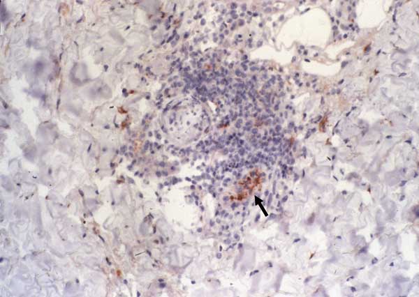
Figure 5. Immunohistochemical detection of Rickettsia africae in the inoculation eschar of a patient with African tick-bite fever. Note the location of the bacteria in the endothelial and inflammatory cells of a blood vessel in the dermis (arrow) (monoclonal rabbit anti-R. africae antibody used at a dilution of 1:1,000 and hematoxylin counterstain; original magnification ×250).
Page created: November 17, 2011
Page updated: November 17, 2011
Page reviewed: November 17, 2011
The conclusions, findings, and opinions expressed by authors contributing to this journal do not necessarily reflect the official position of the U.S. Department of Health and Human Services, the Public Health Service, the Centers for Disease Control and Prevention, or the authors' affiliated institutions. Use of trade names is for identification only and does not imply endorsement by any of the groups named above.