Volume 13, Number 6—June 2007
Research
Levels of Abnormal Prion Protein in Deer and Elk with Chronic Wasting Disease
Figure 1
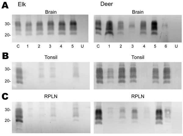
Figure 1. Immunoblot analysis of PrPres from chronic wasting disease (CWD)–affected elk and deer brain, tonsil, and retropharyngeal lymph node (RPLN). Panel A shows the PrPres signal from 2-mg equivalents of elk brain or 20-mg equivalents of deer brain. Individual animals are identified as 1–5 (elk) or 1–6 (deer). C denotes the reference control to which all other samples are compared and consists of 20-mg equivalents of retropharyngeal lymph node (RPLN) from a CWD–affected mule deer. Aliquots of this same control are included on all blots shown in panels B and C. Lanes labeled U in panels A, B, and C contain 20-mg equivalents of the respective tissue from uninfected elk or deer. No PrPres bands were detected when tissues from uninfected deer or elk were analyzed. In panels B and C, 20-mg equivalents of tonsil or RPLN were used. PrPres was obtained as described in Materials and Methods and the blots developed by using antibody L42 at a 0.04 μg/mL dilution and standard enhanced chemifluorescence processing. Approximate molecular weights in kd are indicated on the left side of the panels.