Volume 16, Number 11—November 2010
Letter
Acute Encephalopathy and Pandemic (H1N1) 2009
Song Mi Moon, Sung-Han Kim , Min Hee Jeong, Eun Hye Lee, and Tae-Sung Ko
Figure
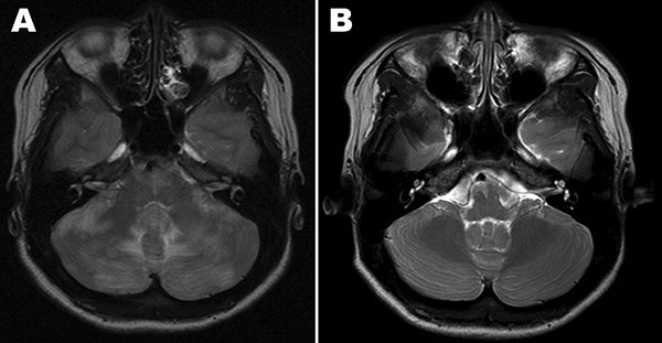
Figure. Magnetic resonance imaging (MRI) scans of case-patient’s brain. A) MRI at hospital admission shows ill-defined T2 changes in both cerebellar hemispheres, periventricular white matter, and the pons. B) MRI of the brain 1 month later, showing nearly complete disappearance of the changes observed at admission.
Page created: March 08, 2011
Page updated: March 08, 2011
Page reviewed: March 08, 2011
The conclusions, findings, and opinions expressed by authors contributing to this journal do not necessarily reflect the official position of the U.S. Department of Health and Human Services, the Public Health Service, the Centers for Disease Control and Prevention, or the authors' affiliated institutions. Use of trade names is for identification only and does not imply endorsement by any of the groups named above.