Skip directly to site content Skip directly to page options Skip directly to A-Z link Skip directly to A-Z link Skip directly to A-Z link
Volume 16, Number 2—February 2010
Dispatch

Pandemic (H1N1) 2009 Outbreak on Pig Farm, Argentina

Ariel Pereda1Comments to Author , Javier Cappuccio1, María A. Quiroga, Elsa Baumeister, Lucas Insarralde, Mariela Ibar, Ramón Sanguinetti, Maria L. Cannilla, Débora Franzese, Oscar E. Escobar Cabrera, Maria I. Craig, Agustina Rimondi, Mariana Machuca, Rosa T. Debenedetti, Carlos Zenobi, Leonardo Barral, Rodrigo Balzano, Santiago Capalbo, Adriana Risso, and Carlos J. Perfumo
Author affiliations: Instituto Nacional de Tecnología Agropecuaria, Castelar, Buenos Aires, Argentina (A. Pereda, M.I. Craig, A. Rimondi); Universidad Nacional de La Plata, La Plata, Buenos Aires (J. Cappuccio, M.A. Quiroga, L. Insarralde, M. Ibar, M. Machuca, C.J. Perfumo); Instituto Nacional de Enfermedades Infecciosas–Administración Nacional de Laboratorios e Institutos de Salud, Buenos Aires (E. Baumeister); Servicio Nacional de Sanidad Animal, Martínez, Buenos Aires (R. Sanguinetti, M.L. Cannilla, D. Franzese, O.E. Escobar Cabrera, R.T. Debenedetti, C. Zenobi, L. Barral, R. Balzano); private veterinary practice, Buenos Aires (S. Capalbo, A. Risso)

Main Article

Figure

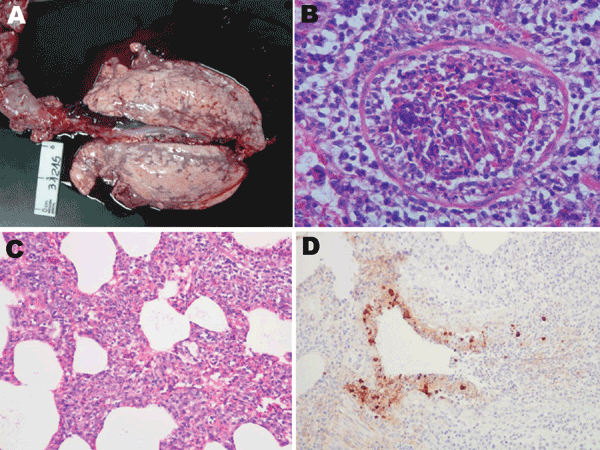
Postmortem samples from clinically affected pigs, Argentina, 2009. A) Macroscopic lung lesions with distinctive scattered dark-red foci of lobular consolidation (chessboard-like) in all lobes. B) Severe necrotizing bronchiolitis with partially denuded epithelia. Hematoxylin-eosin stain; original magnification ×400. C) General view of the alveolar walls showing moderate interstitial thickening by leukocytes. Hematoxin-eosin stain; original magnification ×200. D) Bronchioli. Positive-labeled nucle

Figure. Postmortem samples from clinically affected pigs, Argentina, 2009. A) Macroscopic lung lesions with distinctive scattered dark-red foci of lobular consolidation (chessboard-like) in all lobes. B) Severe necrotizing bronchiolitis with partially denuded epithelia. Hematoxylin-eosin stain; original magnification ×400. C) General view of the alveolar walls showing moderate interstitial thickening by leukocytes. Hematoxin-eosin stain; original magnification ×200. D) Bronchioli. Positive-labeled nuclei and cytoplasm of bronchial epithelial cells and mononuclear cells in the alveoli. 3,3′-diaminobenzidine counterstain with hematoxylin stain; original magnification ×200.

Main Article

1These authors contributed equally to this article.

Page created: May 09, 2011
Page updated: May 09, 2011
Page reviewed: May 09, 2011
The conclusions, findings, and opinions expressed by authors contributing to this journal do not necessarily reflect the official position of the U.S. Department of Health and Human Services, the Public Health Service, the Centers for Disease Control and Prevention, or the authors' affiliated institutions. Use of trade names is for identification only and does not imply endorsement by any of the groups named above.
file_external