Volume 16, Number 7—July 2010
Dispatch
Human Parechovirus Infections in Monkeys with Diarrhea, China
Cite This Article
Citation for Media
Abstract
Information about human parechovirus (HPeV) infection in animals is scant. Using 5′ untranslated region reverse transcription–PCR, we detected HPeV in feces of monkeys with diarrhea and sequenced the complete genome of 1 isolate (SH6). Monkeys may serve as reservoirs for zoonotic HPeV transmissions and as models for studies of HPeV pathogenesis.
Members of the human parechovirus (HPeV) species are small, nonenveloped RNA viruses that are members of the family Picornaviridae, genus Parechovirus. HPeV can be classified into at least 8 genotypes on the basis of sequence similarity of their capsid protein (HPeV-1–HPeV-8). HPeV-1 and HPeV-2, formerly known as echovirus 22 and echovirus 23, were originally considered to belong to the genus Enterovirus (1,2) but after genome sequencing were reclassified as members of a new genus in the family Picornaviridae (3). Recently, 6 other genotypes of parechovirus were isolated from young children with gastrointestinal, respiratory, and severe neurologic signs (4–11). Other HPeV genotypes continue to be characterized (www.picornaviridae.com/parechovirus/hpev/hpev.htm).
Despite the frequent infections and numerous HPeV genotypes detected in humans, information about HPeV infection in animals is scant. In this study, we detected HPeV in feces of monkeys with diarrhea and sequenced the complete genome of 1 isolate (SH6).
In April 2008, fecal specimens were collected from 116 macaques (3–6 years of age) with diarrhea on a monkey farm in People’s Republic of China. Feces were suspended to 10% (wt/vol) in phosphate-buffered saline (0.01 M, pH 7.4), and total RNA was extracted from 200 µL by using TRIZOL reagent (Invitrogen, Carlsbad, CA, USA). Viral RNA was dissolved in 30 µL RNase-free water and stored at –80°C.
Primers (outside-L 5′-CTAGAGAGCTTGGCCGTCGG-3′, outside-R 5′-GTACCTTCTGGGCATCCTTC-3′, inside-L 5′-GGCCTTATACCCCGACTTGC-3′, and inside-R 5′-GGCCTTACAACTAGTGTTTG-3′) (12) were used for reverse transcription nested PCR to identify diverse HPeV genotypes by amplification of a 518-bp fragment located in the 5′ untranslated region (UTR). The expected-size DNA bands were excised from an agarose gel, purified with the AxyPrep DNA gel extraction kit (Axygen, Union City, CA, USA), cloned into pMD-18T vector (TaKaRa, Dalian, China), and sequenced (Applied Biosystems 3730 DNA Analyzer; Invitrogen). Feces from 6 of 116 monkeys were positive for HPeV. The HPeV sequences were compared with those of the HPeV genotype reference strains by using BLAST (www.ncbi.nlm.nih.gov/BLAST). Five of the 6 sequences showed closest identity to the 5′ UTR of HPeV-1 (90%–94%). The viral protein (VP)3/VP1 region of these 5 viruses was then PCR amplified and sequenced to confirm type 1 identity (13). The last monkey feces–derived sequence showed 97%–98% nucleotide identity to HPeV-6 strains. This strain was fully sequenced.
A 674-nt region of the 5′ UTR, an open reading frame (ORF) encoding a polyprotein precursor of 2,182 aa, and a partial 3′ UTR of 88 nt (7,311 bp) were sequenced. The near full genome showed 96% nucleotide identity with the genotype 6 reference genome (AB252582). The polyprotein encoded capsid proteins VP0 (312 aa), VP3 (229 aa), and VP1 (234 aa) and nonstructural proteins 2A (150 aa), 2B (122 aa), 2C (329 aa), 3A (117 aa), 3B (20 aa), 3C (200 aa), and 3D (469 aa). The integrin binding motif arginine–glycine–aspartic acid was identified close to the C terminus of VP1 (8,14).
Figure
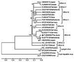
Figure. Phylogenetic analysis of the complete genomes. Phylogenetic tree was constructed by the neighbor-joining method with 1,000 bootstrap replicates using MEGA4.0 software (www.megasoftware.net) with an alignment of the nearly full genome...
We performed phylogenetic analysis using the nearly full genome of SH6 and 17 representative HPeV and related viruses (Figure). Results confirmed that SH6 belonged to genotype 6 and clustered closely with the reference genome from Japan and a strain from the Netherlands (EU077518), forming an HPeV-6 subgroup (Figure).
Previous studies have documented frequent human infections with different HPeV genotypes (www.picornaviridae.com/parechovirus/hpev/hpev.htm). We detected HPeV genotypes 1 and 6 in the feces of 6 of 116 monkeys with diarrhea. A similar analysis of a healthy monkey control group is needed to determine whether an association exists between HPeV infections of monkeys and diarrhea. On the basis of the close similarities between virus feces–derived sequences and HPeV, these viruses might have been transmitted by the fecal–oral route from humans to monkeys. The multiple HPeV genotypes detected indicated that viral transmission occurred on multiple occasions. Monkeys may therefore serve as an animal model for HPeV infection and possibly pathogenesis.
Mr Shan is a PhD candidate at Shanghai Jiao Tong University in Shanghai, China. His interests focus on discovery of novel viruses from biological samples using metagenomic methods.
References
- Joki-Korpela P, Hyypia T. Parechoviruses, a novel group of human picornaviruses. Ann Med. 2001;33:466–71. DOIPubMedGoogle Scholar
- Hyypiä T, Horsnell C, Maaronen M, Khan M, Kalkkinen N, Auvinen P, A distinct picornavirus group identified by sequence analysis. Proc Natl Acad Sci U S A. 1992;89:8847–51. DOIPubMedGoogle Scholar
- Drexler JF, Grywna K, Stöcker A, Almeida PS, Medrado-Ribeiro TC, Eschbach-Bludau M, Novel human parechovirus from Brazil. Emerg Infect Dis. 2009;15:310–3. DOIPubMedGoogle Scholar
- Li L, Victoria J, Kapoor A, Naeem A, Shaukat S, Sharif S, Genomic characterization of novel human parechovirus type. Emerg Infect Dis. 2009;15:288–91. DOIPubMedGoogle Scholar
- Watanabe K, Oie M, Higuchi M, Nishikawa M, Fujii M. Isolation and characterization of novel human parechovirus from clinical samples. Emerg Infect Dis. 2007;13:889–95.PubMedGoogle Scholar
- Benschop KS, Schinkel J, Luken ME, van den Broek PJ, Beersma MF, Menelik N, Fourth human parechovirus serotype. Emerg Infect Dis. 2006;12:1572–5.PubMedGoogle Scholar
- Ito M, Yamashita T, Tsuzuki H, Takeda N, Sakae K. Isolation and identification of a novel human parechovirus. J Gen Virol. 2004;85:391–8. DOIPubMedGoogle Scholar
- Al Sunaidi M, Williams CH, Hughes PJ, Schnurr DP, Stanway G. Analysis of a new human parechovirus allows the definition of parechovirus types and the identification of RNA structural domains. J Virol. 2007;81:1013–21. DOIPubMedGoogle Scholar
- Verboon-Maciolek MA, Groenendaal F, Hahn CD, Hellmann J, van Loon AM, Boivin G, Human parechovirus causes encephalitis with white matter injury in neonates. Ann Neurol. 2008;64:266–73. DOIPubMedGoogle Scholar
- Stanway G, Joki-Korpela P, Hyypia T. Human parechoviruses—biology and clinical significance. Rev Med Virol. 2000;10:57–69. DOIPubMedGoogle Scholar
- Shan TL, Guo W, Cui L, Shang XG, Dai XQ, Yuan CL, The first detection of human parechovirus infections in China. J Clin Virol. 2009;45:371–2. DOIPubMedGoogle Scholar
- Harvala H, Robertson I, McWilliam Leitch EC, Benschop K, Wolthers KC, Templeton K, Epidemiology and clinical associations of human parechovirus respiratory infections. J Clin Microbiol. 2008;46:3446–53. DOIPubMedGoogle Scholar
- Ghazi F, Hughes PJ, Hyypiä T, Stanway G. Molecular analysis of human parechovirus type 2 (formerly echovirus 23). J Gen Virol. 1998;79:2641–50.PubMedGoogle Scholar
Figure
Cite This ArticleTable of Contents – Volume 16, Number 7—July 2010
| EID Search Options |
|---|
|
|
|
|
|
|
Please use the form below to submit correspondence to the authors or contact them at the following address:
X.G. Hua, Building of Agriculture & Biology, Shanghai Jiao Tong University, 800 Dongchuan Rd, Shanghai, 200240, People’s Republic of China
Top