Volume 18, Number 2—February 2012
Dispatch
Plesiomonas shigelloides Infection, Ecuador, 2004–2008
Cite This Article
Citation for Media
Abstract
Diarrheal risk associated with Plesiomonas shigelloides infection was assessed in rural communities in northwestern Ecuador during 2004–2008. We found little evidence that single infection with P. shigelloides is associated with diarrhea but stronger evidence that co-infection with rotavirus causes diarrhea.
Plesiomonas shigelloides (family Enterobacteriaceae) has been implicated in gastroenteritis outbreaks in travelers to tropical regions and in persons who have ingested contaminated food or water (1–3). For persons native to tropical regions, however, case–control studies have found little or no association between P. shigelloides infection and diarrhea (4–6). Although these studies have been conducted in areas where mixed infections are generally common, to our knowledge, none examined co-infections. We assessed the pathogenicity of P. shigelloides in the context of co-infections and across all age groups in a province in northwestern Ecuador.
During 2004–2008, serial case–control studies were conducted in 22 remote communities in Esmeraldas Province, Ecuador. Complete study design and laboratory procedures for pathogen detection have been described (7). Briefly, each community was visited 4–6 times on a rotating basis; each visit lasted for 15 days, during which all cases of diarrhea were identified by a visit to each household every morning. Household residents with cases had >3 loose stools in a 24-hour period, and controls had no symptoms of diarrhea during the past 6 days. Fecal samples were collected from 3 healthy controls per person with diarrhea. These samples were plated on selective agar media, and 5 lactose-fermenting colonies were screened by PCR for enterotoxigenic Escherichia coli (ETEC), enteropathogenic E. coli, and enteroinvasive E. coli (EIEC). Lactose-negative isolates that were identified as either Shigella spp. or E. coli were also screened by PCR for the same molecular marker used for EIEC. All non–lactose-fermenting pathogens, including P. shigelloides, were biochemically identified by API 20E system (bioMèrieux, Marey l’Etoile, France). Because shigellae are phylogenetically similar to E. coli pathotypes, we combined data from persons infected with E. coli and those infected with shigellae in our analysis. We tested for Giardia lamblia by using an ELISA kit (RIDASCREEN Giardia; R-Biopharm, Darmstadt, Germany), and rotavirus was detected with an enzyme immunoassay kit (RIDA Quick Rotavirus; R-Biopharm). We chose a molecular method for detecting E. coli pathotypes because they cannot be differentiated solely on the basis of biochemical tests; the metabolic homogeneity of P. shigelloides, however, makes this organism easily and clearly identifiable by biochemical test. Similarly, immunologic methods used for Giardia spp. and rotavirus detection are specific and sensitive enough to accurately detect these pathogens, and use of molecular methods would be justified only for deeper analysis. Institutional review board committees at the University of California, Berkeley; University of Michigan; Trinity College; and Universidad San Francisco de Quito approved all protocols.
Figure
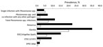
Figure. Case prevalence (black) and weighted community prevalence (white) of enteric pathogens, Ecuador, 2004–2008. Identification of pathogenic Escherichia coli was based on the genes given in parentheses. EIEC, enteroinvasive E. coli; Ipah,...
During March 2004–March 2008, a total of 2,936 fecal samples were collected from persons of all ages (168 [6%] were <1 year of age, 597 [20%] were 1–4 years, 753 [26%] were 5–12 years, 1,362 [46%] were >13 years, and 56 [2%] were missing a birth date), corresponding to 775 cases and 2,161 controls. P. shigelloides was isolated in 253 (8.6%) samples. This number exceeded isolation rates for all of the pathogens analyzed except G. lamblia, which was present in 701 (23.9%) samples. Rotavirus was detected in 225 (7.7%) samples and EIEC/shigellae in 188 (6.4%) samples. P. shigelloides was detected in 11.4% of case-patients with diarrhea (case prevalence), which is more than the 7.2% estimated in the community (weighted control prevalence; Figure). However, once we stratified by persons infected only with P. shigelloides and those infected with P. shigelloides plus >1 of the other marker pathogens for which we tested, single infections with P. shigelloides were almost equally prevalent in the case-patients and in the community; in contrast, co-infections with P. shigelloides and other pathogens were more frequent in persons with diarrhea (Figure).
To determine whether P. shigelloides infection was associated with diarrhea, we estimated risk ratios (RRs) and bootstrapped 95% CIs for single and co-infection exposures (Table). A single infection with P. shigelloides was not associated with diarrhea (RR 1.5, 95% CI 0.9–2.2). Persons co-infected with P. shigelloides and another pathogen, however, had almost 6× the risk for diarrhea than those with no infection (RR 5.6, 95% CI 3.5–9.3) and simultaneous occurrence of P. shigelloides and rotavirus increased the risk for diarrhea to 16.2 (Table). We found no evidence for confounding of the association between P. shigelloides and diarrhea by co-infecting pathogens (RRcrude = RRMH-pooled; where RRcrude is the unadjusted RR and RRMH-pooled is the pooled Mantel-Haenszel RR estimate). However, we found some evidence for confounding by age of P. shigelloides co-infection (RRMH-pooled 4.2 [95% CI 2.1–8.1], compared with the crude estimate of 5.6) but no evidence for confounding by age for single infection with P. shigelloides.
A single infection with P. shigelloides resulted in a moderately increased risk for diarrheal disease, which suggests that this microorganism plays a minor role as a pathogen. This result agrees with findings of previous studies (4,8,9). Analysis of the co-infections, however, suggests that P. shigelloides may be pathogenic in the presence of another pathogen. Specifically, co-infections of P. shigelloides with either rotavirus or pathogenic E. coli (including shigellae) were 16.2× (95% CI 5.5–62.3) and 13.8× (95% CI 3.3–69.3) more likely to result in diarrhea, respectively. We cannot, therefore, rule out the pathogenic capacity of P. shigelloides even though single infection may not be sufficient to cause disease.
This co-infection analysis might be limited by the number of pathogens considered (Giardia spp., rotavirus, pathogenic E. coli, and shigellae). However, the high isolation rates suggest we are detecting the major pathogens in the region. Other pathogens that may be useful to consider, given their attention in the literature, include Entaemobae histolytica and Cryptosporidium spp.
Although we found nothing in the literature that addresses the role of co-infection in the pathogenicity of P. shigelloides, co-infection with enteric pathogens is a well-known phenomenon, especially in tropical regions (6). Co-infection with ETEC and enteropathogenic E. coli increases virulence (10). Other studies have shown that the severity of disease is increased when rotavirus infections occur alongside another infection with another enteric pathogen (11).
P. shigelloides may take advantage of the disruption of the normal gut microbiota and gut physiology because of the concurrent presence of other pathogens, establishing a pathology in the human gut. For example, diarrhea caused by enterotoxins produced by pathogens, such as ETEC, and Vibrio cholerae (12), may remove normal gut microbiota, enabling P. shigelloides to establish an infection. The disruption of gut microbiota that facilitates gut colonization has been demonstrated in murine models infected with Citrobacter rodentium and Salmonella enterica serovar Typhimurium (13).
Most medical literature considers infectious diarrhea as a monopathogenic phenomenon (12,14). In the data presented here, the crude risk ratio suggests that P. shigelloides is pathogenic. When looking at single infections, we found no evidence that P. shigelloides is pathogenic. When looking at co-infection data, however, we found associations between infection and diarrhea. Our findings suggest that multipathogenic infections may play a role in the pathogenesis of infectious diarrhea.
Dr Escobar is a graduate student at Universidad San Francisco de Quito, Quito, Ecuador. His research interests are molecular epidemiology of enteric pathogens and co-infections that cause diarrheal diseases.
Acknowledgment
This study was supported by the US National Institute of Allergy and Infectious Disease, grant no. RO1-AI050038.
References
- Adams MR, Moss MO. Food microbiology. 3rd ed. Cambridge (UK): The Royal Society of Chemistry; 2008.
- Brenden RA, Miller MA, Janda JM. Clinical disease spectrum and pathogenic factors associated with Plesiomonas shigelloides infections in humans. Rev Infect Dis. 1988;10:303–16. DOIPubMedGoogle Scholar
- Kain KC, Kelly MT. Clinical features, epidemiology, and treatment of Plesiomonas shigelloides diarrhea. J Clin Microbiol. 1989;27:998–1001.PubMedGoogle Scholar
- Pitarangsi C, Echeverria P, Whitmire R, Tirapat C, Formal S, Dammin GJ, Enteropathogenicity of Aeromonas hydrophila and Plesiomonas shigelloides: prevalence among individuals with and without diarrhea in Thailand. Infect Immun. 1982;35:666–73.PubMedGoogle Scholar
- Alabi SA, Odugbemi T. Occurrence of Aeromonas species and Plesiomonas shigelloides in patients with and without diarrhoea in Lagos, Nigeria. J Med Microbiol. 1990;32:45–8. DOIPubMedGoogle Scholar
- Bodhidatta L, McDaniel P, Sornsakrin S, Srijan A, Serichantalergs O, Mason CJ. Case–control study of diarrheal disease etiology in a remote rural area in western Thailand. Am J Trop Med Hyg. 2010;83:1106–9. DOIPubMedGoogle Scholar
- Eisenberg JN, Cevallos W, Ponce K, Levy K, Bates SJ, Scott JC, Environmental change and infectious disease: how new roads affect the transmission of diarrheal pathogens in rural Ecuador. Proc Natl Acad Sci U S A. 2006;103:19460–5. DOIPubMedGoogle Scholar
- Abbott SL, Kokka RP, Janda JM. Laboratory investigations on the low pathogenic potential of Plesiomonas shigelloides. J Clin Microbiol. 1991;29:148–53.PubMedGoogle Scholar
- Albert MJ, Faruque AS, Faruque SM, Sack RB, Mahalanabis D. Case–control study of enteropathogens associated with childhood diarrhea in Dhaka, Bangladesh. J Clin Microbiol. 1999;37:3458–64.PubMedGoogle Scholar
- Crane JK, Choudhari SS, Naeher TM, Duffey ME. Mutual enhancement of virulence by enterotoxigenic and enteropathogenic Escherichia coli. Infect Immun. 2006;74:1505–15. DOIPubMedGoogle Scholar
- Grimprel E, Rodrigo C, Desselberger U. Rotavirus disease: impact of coinfections. Pediatr Infect Dis J. 2008;27:S3–10. DOIGoogle Scholar
- Sussman M, editor. Molecular medical microbiology. London: Academic Press; 2002.
- Viswanathan VK, Hodges K, Hecht G. Enteric infection meets intestinal function: how bacterial pathogens cause diarrhoea. Nat Rev Microbiol. 2009;7:110–9.PubMedGoogle Scholar
- Guandalini S, Vaziri H, eds. Diarrhea: diagnostic and therapeutic advances. London: Springer; 2011.
Figure
Table
Cite This ArticleTable of Contents – Volume 18, Number 2—February 2012
| EID Search Options |
|---|
|
|
|
|
|
|
Please use the form below to submit correspondence to the authors or contact them at the following address:
Joseph Eisenberg, University of Michigan, 1415 Washington Heights, Ann Arbor, MI 48109, USA
Top