Volume 18, Number 8—August 2012
Dispatch
New Variants of Porcine Epidemic Diarrhea Virus, China, 2011
Figure 1
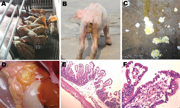
Figure 1. . Clinical features of pigs infected with porcine epidemic diarrhea virus from pig farms in the People’s Republic of China, 2011. A) Litter of pigs infected with this virus, showing watery diarrhea and emaciated bodies. B) A representative emaciated piglet with yellow, water-like feces. C) Yellow and white vomitus from a representative sucking piglet. D) Thin-walled intestinal structure with light yellow water-like content. E) Congestion in the small intestinal wall and intestinal villi; desquamated epithelial cells from the intestinal villus (original magnification ×100). F) Congestion in the lamina propria of intestinal mucosa, and degeneration, necrosis, and desquamation of epithelial cells of the intestinal villi (original magnification ×400).