Volume 19, Number 1—January 2013
Letter
Schmallenberg Virus in Central Nervous System of Ruminants
Cite This Article
Citation for Media
To the Editor: In 2011, a new virus spread throughout ruminant populations in the Netherlands, Germany, and other European countries (1). Infected dairy cattle exhibited fever, reduced milk yield, loss of appetite, and diarrhea. Weeks after these signs appeared, epidemic abortions; births of malformed or stillborn animals; and perinatal deaths of calves, lambs, and goat kids were reported. Metagenomic analysis identified a novel orthobunyavirus (family Bunyaviridae), termed Schmallenberg virus (SBV), which is closely related to Akabane and Shamonda viruses (1). These arthropod-borne viruses are well-recognized ruminant pathogens in Africa, Asia, and Oceania; fetal infection is associated with an arthrogryposis–hydranencephaly syndrome.
Previous reports about SBV have addressed clinical signs (2), serologic findings (3), pathologic findings (4), and SBV RNA distribution in organs of malformed calves (5) detected by real-time quantitative reverse transcription PCR (qRT-PCR). However, pathogenesis and viral cell tropism are largely unknown. Therefore, our aims were to establish an in situ hybridization method (ISH) to detect SBV mRNA, to evaluate the usefulness of this method as a complementary diagnostic tool, and to further analyze SBV pathogenesis.
The in situ probe was generated according to established protocols (6). The SBV qRT-PCR product and primers used for diagnostics (5) were used to amplify an 88-bp segment of the nucleoprotein, encoded by the small segment of the SBV genome. The amplicon was cloned into a pCR4-TOPO vector (Invitrogen, Darmstadt, Germany) and sequenced. For generation of a digoxigenin-labeled antisense probe to detect viral mRNA, we used M13 reverse and SBV-S-469R primers (5). Specificity of the probe was ensured by distinct ISH signals in the brains of SBV-positive (as determined by qRT-PCR) animals (6). No reaction was detected in SBV-negative (as determined by qRT-PCR) goats, sheep, and calves; in ruminants with various non–SBV-associated nervous system lesions; or in the brain of a mouse that was experimentally infected with Akabane virus.
Thereafter, we investigated SBV mRNA distribution in the central nervous system (CNS) of 82 naturally infected ruminants (46 lambs, 2 goat kids, and 34 calves), all of which had previously been found by qRT-PCR to be positive for SBV. In addition, ISH was used to examine the following tissues from 10 of these animals (4 lambs, 1 goat kid, and 5 calves): placenta, muscle, eye, heart, aorta, lung, trachea, liver, kidney, spleen, small and large intestine, mesenteric and pulmonary lymph nodes, thymus, adrenal gland, testis, and uterus.
Figure
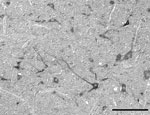
SBV mRNA was found in varying amounts, predominantly in neurons (Figure) of the cerebrum, cerebellum, brain stem, medulla oblongata, and spinal cord of 7 lambs, 1 goat kid, and 2 calves. Randomly distributed clusters of SBV-positive neurons were frequently found in small ruminants, whereas only single positive cells were found in both calves. SBV mRNA was not found in any peripheral organ. The Technical Appendix provides an overview of ISH results in relation to malformations and inflammatory CNS lesions. Histologic examination identified encephalitis, characterized by lymphohistiocytic, perivascular cuffs, in 11 (9 lambs, 1 goat kid, and 1 calf) of the 82 animals. Among animals that were positive according to ISH, all small ruminants showed inflammation, whereas both calves lacked inflammatory changes.
The most frequent macroscopic lesions or conditions in animals that were SBV positive by qRT-PCR, especially calves, were arthrogryposis, brachygnathia inferior, torticollis, kyphosis, lordosis, scoliosis, and muscle hypoplasia (especially in calves) (4). The predominant CNS lesions or conditions were cerebellar and cerebral hypoplasia, hydranencephaly, porencephaly, hydrocephalus, and micromyelia. Among the 8 small ruminants that were SBV positive by ISH, hydrocephalus was found in 5, cerebellar hypoplasia in 5, hydranencephaly in 1, and arthrogryposis in 6. For the 2 calves that were SBV positive, micromyelia and arthrogryposis were found in both and cerebellar hypoplasia in 1.
This study indicates that neurons are the predominant target in SBV-infected neonates. Surprisingly, only 10 of 82 ruminants were positive for SBV by qRT-PCR and ISH. This discrepancy could result from low SBV mRNA copy numbers per cell, which are detectable by qRT-PCR but not ISH, and/or from reduced viral load in individual cells because of the long time between assumed infection in early gestation and time of examination after birth (≈17 weeks in sheep and 27 weeks in calves). The latter explanation is indicated by the high incidence of malformations (7) typical for teratogenic insults and might especially apply to calves. Detection of CNS inflammation in all small ruminants that were SBV positive according to ISH indicates a cause-and-effect association between high SBV mRNA copy numbers per cell and lymphohistiocytic immune response. Furthermore, low numbers of infected cells and/or reduced replication as assumed for animals that were positive by qRT-PCR but negative by ISH might explain the absence of inflammation in the calf brains. The 3 tested animals in which inflammation was found but that were SBV negative by ISH could represent an advanced resolving lesion after viral clearance or reduction. In situ detection of SBV mRNA represents a suitable way to study SBV pathogenesis, especially in the active phase of infection, and might enable identification of SBV as the causative agent in cases of CNS inflammation of unknown etiology.
Acknowledgment
We thank Takafumi Hamaoka for providing the brain samples of the Akabane virus–infected mouse and Danuta Waschke, Bettina Buck, Caroline Schütz, and Claudia Herrmann for excellent technical assistance.
References
- Hoffmann B, Scheuch M, Höper D, Jungblut R, Holsteg M, Schirrmeier H, Novel orthobunyavirus in cattle, Europe, 2011. Emerg Infect Dis. 2012;18:469–72. DOIPubMedGoogle Scholar
- Gariglinany M-M, Hoffmann B, Dive M, Sartelet A, Bayrou C, Cassart D, Schmallenberg virus in calf born at term with porencephaly, Belgium. Emerg Infect Dis. 2012;18:1005–6. DOIGoogle Scholar
- Elbers AR, Loeffen WL, Quak S, de Boer-Luijtze E, van der Spek AN, Bouwstra R, Seroprevalence of Schmallenberg virus antibodies among dairy cattle, the Netherlands, winter 2011–2012. Emerg Infect Dis. . Epub 2012 May.DOIPubMedGoogle Scholar
- Herder V, Wohlsein P, Peters M, Hansmann F, Baumgärtner W. Salient lesions in domestic ruminants infected with the emerging so-called Schmallenberg virus in Germany. Vet Pathol. 2012;49:588–91. DOIPubMedGoogle Scholar
- Bilk S, Schulze C, Fischer M, Beer M, Hlinak A, Hoffmann B. Organ distribution of Schmallenberg virus RNA in malformed newborns. Vet Microbiol. . Epub 2012 March 30.DOIPubMedGoogle Scholar
- Gröters S, Alldinger S, Baumgärtner W. Up-regulation of mRNA for matrix metalloproteinases-9 and -14 in advanced lesions of demyelinating canine distemper leukoencephalitis. Acta Neuropathol. 2005;110:369–82. DOIPubMedGoogle Scholar
Figure
Cite This Article1These authors contributed equally to this article.
Related Links
Table of Contents – Volume 19, Number 1—January 2013
| EID Search Options |
|---|
|
|
|
|
|
|
Please use the form below to submit correspondence to the authors or contact them at the following address:
Wolfgang Baumgärtner, Department of Pathology, University of Veterinary Medicine, Bünteweg 17, 30559 Hannover, Germany
Top