Volume 19, Number 2—February 2013
CME ACTIVITY - Research
Laboratory-based Surveillance for Hepatitis E Virus Infection, United States, 2005–2012
Cite This Article
Citation for Media
Introduction
![]()
Medscape, LLC is pleased to provide online continuing medical education (
This activity has been planned and implemented in accordance with the Essential Areas and policies of the Accreditation Council for Continuing Medical Education through the joint sponsorship of Medscape, LLC and Emerging Infectious Diseases. Medscape, LLC is accredited by the ACCME to provide continuing medical education for physicians.
Medscape, LLC designates this Journal-based CME activity for a maximum of 1 AMA PRA Category 1 Credit(s)TM. Physicians should claim only the credit commensurate with the extent of their participation in the activity.
All other clinicians completing this activity will be issued a certificate of participation. To participate in this journal CME activity: (1) review the learning objectives and author disclosures; (2) study the education content; (3) take the post-test with a 70% minimum passing score and complete the evaluation at http://www.medscape.org/journal/eid; (4) view/print certificate.
Release date: Release date: January 23, 2013; Expiration date: January 23, 2014
Learning Objectives
Upon completion of this activity, participants will be able to:
• Describe the percentage of hepatitis E among US patients with hepatitis who were seronegative for acute hepatitis A and B, including those who had and those who had not traveled abroad
• Compare characteristics of nontravelers vs travelers with hepatitis E
• Describe HEV genotypes among nontravelers vs travelers with hepatitis E.
CME Editor
Shannon O’Connor, ELS, Technical Writer/Editor, Emerging Infectious Diseases. Disclosure: Shannon O’Connor has disclosed no relevant financial relationships.
CME Author
Laurie Barclay, MD, freelance writer and reviewer, Medscape, LLC. Disclosure: Laurie Barclay, MD, has disclosed no relevant financial relationships.
Authors
Disclosures: Jan Drobeniuc; Tracy Greene-Montfort; Le Ngoc-Thao; Tonya R. Mixson-Hayden; Lilia Ganova-Raeva; Chen Dong; Ryan T. Novak; Umid M. Sharapov; Rania A. Tohme; Eyasu Teshale; Saleem Kamili; and Chong-Gee Teo have disclosed no relevant financial relationships.
Abstract
To investigate characteristics of hepatitis E cases in the United States, we tested samples from persons seronegative for acute hepatitis A and B whose clinical specimens were referred to the Centers for Disease Control and Prevention during June 2005–March 2012 for hepatitis E virus (HEV) testing. We found that 26 (17%) of 154 persons tested had hepatitis E. Of these, 15 had not recently traveled abroad (nontravelers), and 11 had (travelers). Compared with travelers, nontravelers were older (median 61 vs. 32 years of age) and more likely to be anicteric (53% vs. 8%); the nontraveler group also had fewer persons of South Asian ethnicity (7% vs. 73%) and more solid-organ transplant recipients (47% vs. 0). HEV genotype 3 was characterized from 8 nontravelers and genotypes 1 or 4 from 4 travelers. Clinicians should consider HEV infection in the differential diagnosis of hepatitis, regardless of patient travel history.
Hepatitis E in Africa, southern and central Asia, and Central America causes occasional outbreaks of jaundice and, between outbreaks, occurrences of sporadic jaundice. Primarily spread by waterborne transmission, the disease tends to resolve spontaneously, although fulminant hepatic failure can ensue (1). In eastern Asia and Europe, sporadic hepatitis E, whether imported after return from international travel or acquired indigenously, has been observed; the indigenous form is thought to be foodborne (2). Although the disease is largely self-limiting, in Europe, chronic hepatitis E, which may lead to cirrhosis, is increasingly recognized among solid-organ transplant recipients (SOTRs) (3).
The causative agent of hepatitis E is hepatitis E virus (HEV), of which 4 genotypes are found in humans. Genotypes 1 and 2 circulate in regions where waterborne transmission is common; genotype 3 is prevalent in eastern Asia and the West and genotype 4 in eastern Asia. Genotypes 1 and 2 infect humans, but genotypes 3 and 4 infect humans and animals, predominantly pigs (4).
In the United States, HEV imported into the country after travel to regions to which waterborne HEV transmission is endemic is well recognized (5–7). Recently, 21% of participants of the US-based Third National Health and Nutrition Examination Survey were found seropositive for IgG against HEV (8). This unexpectedly high prevalence rate would not be ascribable to imported HEV infection alone. Indeed, cases of hepatitis E unassociated with travel abroad have been observed in the United States, implying infection by indigenous HEV strains (9–14). Moreover, the increasing number of reports from Europe of hepatitis E among SOTRs (3,15) suggests that SOTRS in the United States might be similarly susceptible to the disease.
We report a study of demographic, clinical, travel-related, and virologic characteristics of persons with hepatitis E derived from a diverse patient base. Critical to this investigation was the application of a validated serologic assay for detecting IgM against HEV (16), the marker of recent HEV infection, as well as a real-time reverse transcription PCR (RT-PCR) that had been validated to detect, to high sensitivity, HEV RNA (17), which is an indicator of active HEV shedding. Together, these 2 assays enabled us to identify patients with incident hepatitis E.
Samples and Patients
The Centers for Disease Control and Prevention (CDC) conducts HEV testing of serum and stool samples referred by health care providers, public health departments, and diagnostic laboratories in the United States (18). Referrers are requested to fill out a standardized questionnaire of patients’ demographics, clinical and laboratory test features, and risks for HEV infection, including recent international travel and destinations visited; the completed questionnaire is submitted along with the test specimens (18). Persons whose specimens were received during June 2005–March 2012 and reported as being negative for IgM against hepatitis A virus and hepatitis B core antigen, regardless of positivity for IgG against hepatitis C virus, were considered for inclusion into the study.
Assays
An earlier, pangenotypic evaluation by CDC of 6 serologic assays for IgM against HEV identified the assay manufactured by Diagnostic Systems (Saronno, Italy) as having the best performance characteristics (16). Its diagnostic sensitivity and specificity were 98% and 95.2%, respectively, and its analytic sensitivity was 9 Walter Reed Units/mL. For this study, the assay was used to detect IgM against HEV in test samples. IgG against HEV was tested by applying an assay from the same manufacturer. Serum and stool samples were tested for HEV RNA by a real-time RT-PCR, capable of detecting HEV genotypes 1–4 to a sensitivity of 4 HEV genome-equivalents/mL, to amplify a 69-bp fragment in open reading frame (ORF) 3 of the HEV genome (19). Application of that assay enabled our laboratory to attain perfect detection scores in a recent international evaluation of 20 laboratories conducting HEV RNA testing (17). Samples found to be positive for HEV RNA were subjected to another RT-PCR to generate amplicons from a 258-bp segment from ORF1, which were then processed for nucleotide sequencing and phylogenetic analyses (20).
Statistics
Distributions of variables were assessed by using the Kruskal-Wallis test and the χ2 test with the Yates correction or the Fisher exact test, as appropriate. Univariate and bivariate data analyses were conducted by using Epi Info (wwwn.cdc.gov/EpiInfo/html/prevVersion.htm).
Case Definition
A case of hepatitis E was defined as illness in a person in whom IgM and IgG against HEV in serum or HEV RNA in serum or stool samples were detected. A person in whom IgM but not IgG against HEV was detected in serum was excluded unless HEV RNA was found or IgG againstHEV was detected in follow-up serum samples. A person in whom IgG but not IgM against HEV was detected in serum samples was included if HEV RNA was found in serum or stool samples.
Of 154 persons whose specimens fulfilled the inclusion criteria, 26 (17%) met the case definition for hepatitis E. Case-patients were between 14–67 years of age (median 43 years); 19 (73%) were male. Fifteen (58%) were white, 9 (36%) South Asian, and 2 (8%) Hispanic. None were seropositive for IgG against HEV. Eighteen (69%) case-patients were jaundiced, and 7 (27%) were SOTRs, the allografts received being kidney (3), liver (2), kidney and pancreas (1), and heart and lungs (1). Fifteen case-patients (58%) who reported not having traveled outside the United States in the previous 2 months were classified as nontravelers; the remaining 11, who had traveled abroad, were classified as travelers.
The Table summarizes the demographic, clinical, and virologic data for individual case-patients. Compared with travelers, nontravelers were older (median age 61 vs. 32 years of age; p<0.05) and more likely to be anicteric (not jaundiced; 8/15 [53%] vs. 1/11 [8%]; p = 0.02). The nontraveler group also included fewer South Asians (1/15 [7%] vs. 8/11 [73%]; p<0.001) and more SOTRs (7/15 [47%] vs. 0; p = 0.02). Differences in sex distribution were not significant.
Three case-patients (NT6, NT7, and T9; Table) were documented to have fulminant hepatic failure. Two of them required liver transplantation; 1 died and 1 survived.
Figure
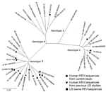
Figure. . . . Genetic relatedness among hepatitis E virus (HEV) strains identified in hepatitis E cases, United States. Phylogenetic tree was constructed from a segment of HEV open reading frame...
HEV RNA was amplified from 12 case-patients (46%). The rate of HEV RNA detection among SOTRs (5/7; 71%) was higher than that among non-SOTRs (7/19; 37%), but this difference was not significant. HEV genotype 1 was characterized from 3 travelers, genotype 3 from 8 nontravelers (including the 5 SOTRs), and genotype 4 from 1 traveler. The Figure displays the genetic diversity of HEV carried.
We identified 26 case-patients with hepatitis E in the United States. No distinction was made between acute and chronic hepatitis E. Whereas acute hepatitis among non-SOTRs was readily identifiable (most were jaundiced when test specimens were drawn), it was difficult to asses whether disease in SOTRs was at the acute or chronic stage during specimen sampling, because positivity for IgM against HEV or HEV RNA could reflect either stage (3). Thus, the case definition was kept broad to identify both stages of disease. As the study was not primarily prospective, the natural history of hepatitis E among the case-patients was largely unknown. Nevertheless, adverse outcomes could be documented for 3 case-patients, in whom fulminant hepatic failure developed.
Hepatitis E cases were found among persons who had not recently traveled abroad and those who had. Nontravelers tended to be older than travelers, a trend consistent with the finding recently reported by the Drug-Induced Liver Injury Network of 9 patients seropositive for IgM against HEV whose mean age was 67 years (21) and with similar observations in Japan and Europe (1,2,22). The higher proportion of anicteric persons in the nontraveler group reflects its inclusion of all SOTRs, which in Europe have been observed to have largely asymptomatic infections (3).
Nontravelers were infected exclusively by HEV genotype 3 strains. These strains clustered with HEV previously found in case-patients with nonimported acute hepatitis E (9–12,14) in the United States (Figure), suggesting that the nontravelers were infected by autochthonously circulating HEV. The similarity between HEV genotype 3 strains identified in nontravelers with those in swine (4) (Figure) suggests, but does not prove, HEV transmission linkage between humans and pigs (2). Evidence of HEV infection acquired after consumption of inadequately cooked meat and offal originating from pigs, boars, and deer has been reported from Japan (2) and France (23). Elsewhere, including the United States, evidence implicating non–travel-associated hepatitis E as a zoonosis remains weak (24).
The patient base from which hepatitis E cases were identified was nonselective, broad, and derived from multiple health care provider contexts. Nonetheless, data from this study were not generated from an established, systematic program of epidemiologic surveillance. Accordingly, the cases identified here may not fully represent the extent of hepatitis E in the United States. The larger number of cases among nontravelers likely reflects more persons living in the United States who do not travel abroad compared with those who do, and the many SOTRs identified with hepatitis E could be an overrepresentation resulting from increasing awareness among physicians of the predilection of the SOTR patient subpopulation to HEV infection (3,15). Future surveillance of hepatitis E may need to sample source populations from more diverse settings, such as gastroenterology/hepatology clinics (21), travel clinics (6), and the military (7). We recently reported findings from a study of HEV infection among immunocompromised patients other than SOTRs (25).
This study has provided insight into nonimported and imported hepatitis E in the United States. The nonimported form was observed to affect SOTRs, be able to lead to adverse outcomes, and be associated with infection by HEV genotype 3. The extent of nonimported hepatitis E in the United States merits further investigation, as does the role of autochthonous transmission of genotype 3 HEV strains. In clinical practice, entry of hepatitis E into the differential diagnosis of suspected hepatitis, regardless of the patient’s travel history, would be appropriate.
Dr Drobeniuc is a microbiologist in the Division of Viral Hepatitis, Centers for Disease Control and Prevention. His research interests include laboratory diagnostics, assay development, quality assurance, and epidemiology relating to viral hepatitis.
References
- Labrique A, Kuniholm MH, Nelson KE. The global impact of hepatitis E—new horizons for an emerging virus. In: Grayson L, editor. Emerging Infections 9. Washington (DC): American Society for Microbiology; 2010. p. 54–92.
- Teo CG. Much meat, much malady: changing perceptions of the epidemiology of hepatitis E. Clin Microbiol Infect. 2010;16:24–32. DOIPubMedGoogle Scholar
- Legrand-Abravanel F, Kamar N, Sandres-Saune K, Garrouste C, Dubois M, Mansuy JM, Characteristics of autochthonous hepatitis E virus infection in solid-organ transplant recipients in France. J Infect Dis. 2010;202:835–44. DOIPubMedGoogle Scholar
- Dong C, Meng J, Dai X, Liang JH, Feagins AR, Meng XJ, Restricted enzooticity of hepatitis E virus genotypes 1 to 4 in the United States. J Clin Microbiol. 2011;49:4164–72. DOIPubMedGoogle Scholar
- Centers for Disease Control and Prevention. Hepatitis E among U.S. travelers, 1989–1992. MMWR Morb Mortal Wkly Rep. 1993;42:1–4 .PubMedGoogle Scholar
- Ooi WW, Gawoski JM, Yarbough PO, Pankey GA. Hepatitis E seroconversion in United States travelers abroad. Am J Trop Med Hyg. 1999;61:822–4 .PubMedGoogle Scholar
- Eick A, Ticehurst J, Tobler S, Nevin R, Lindler L, Hu Z, Hepatitis E seroprevalence and seroconversion among US military service members deployed to Afghanistan. J Infect Dis. 2010;202:1302–8. DOIPubMedGoogle Scholar
- Kuniholm MH, Purcell RH, McQuillan GM, Engle RE, Wasley A, Nelson KE. Epidemiology of hepatitis E virus in the United States: results from the Third National Health and Nutrition Examination Survey, 1988–1994. J Infect Dis. 2009;200:48–56. DOIPubMedGoogle Scholar
- Kwo PY, Schlauder GG, Carpenter HA, Murphy PJ, Rosenblatt JE, Dawson GJ, Acute hepatitis E by a new isolate acquired in the United States. Mayo Clin Proc. 1997;72:1133–6. DOIPubMedGoogle Scholar
- Erker JC, Desai SM, Schlauder GG, Dawson GJ, Mushahwar IK. A hepatitis E virus variant from the United States: molecular characterization and transmission in cynomolgus macaques. J Gen Virol. 1999;80:681–90 .PubMedGoogle Scholar
- Tsang TH, Denison EK, Williams HV, Venczel LV, Ginsberg MM, Vugia DJ. Acute hepatitis E infection acquired in California. Clin Infect Dis. 2000;30:618–9. DOIPubMedGoogle Scholar
- Amon JJ, Drobeniuc J, Bower WA, Magaña JC, Escobedo MA, Williams IT, Locally acquired hepatitis E virus infection, El Paso, Texas. J Med Virol. 2006;78:741–6. DOIPubMedGoogle Scholar
- Curry JA, Adams N, Crum-Cianflone NF. Acute hepatitis E virus infection in an HIV-infected person in the United States. Ann Intern Med. 2009;150:226–7 .PubMedGoogle Scholar
- Tohme RA, Drobeniuc J, Sanches R, Heseltine G, Alsip B, Kamili S, Acute hepatitis associated with autochthonous hepatitis E virus infection — San Antonio, Texas, 2009. Clin Infect Dis. 2011;53:793–6. DOIPubMedGoogle Scholar
- Pas SD, de Man RA, Mulders C, Balk AH, van Hal PT, Weimar W, Hepatitis E virus infection among solid organ transplant recipients, the Netherlands. Emerg Infect Dis. 2012;18:869–72. DOIPubMedGoogle Scholar
- Drobeniuc J, Meng J, Reuter G, Greene-Montfort T, Khudyakova N, Dimitrova Z, Serologic assays specific to immunoglobulin M antibodies against hepatitis E virus: pangenotypic evaluation of performances. Clin Infect Dis. 2010;51:e24–7. DOIPubMedGoogle Scholar
- Baylis SA, Hanschmann KM, Blümel J, Nübling CM; HEV Collaborative Study Group. Standardization of hepatitis E virus (HEV) nucleic acid amplification technique–based assays: an initial study to evaluate a panel of HEV strains and investigate laboratory performance. J Clin Microbiol. 2011;49:1234–9. DOIPubMedGoogle Scholar
- Centers for Disease Control and Prevention. Hepatitis E information for healthcare professionals [cited 2012 Sep 14]. http://www.cdc.gov/hepatitis/HEV/LabTestingRequests.htm
- Jothikumar N, Cromeans TL, Robertson BH, Meng XJ, Hill VR. A broadly reactive one-step real-time RT-PCR assay for rapid and sensitive detection of hepatitis E virus. J Virol Methods. 2006;131:65–71. DOIPubMedGoogle Scholar
- Chatterjee R, Tsarev S, Pillot J, Coursaget P, Emerson SU, Purcell RH. African strains of hepatitis E virus that are distinct from Asian strains. J Med Virol. 1997;53:139–44. DOIPubMedGoogle Scholar
- Davern TJ, Chalasani N, Fontana RJ, Hayashi PH, Protiva P, Kleiner DE, ; Drug-Induced Liver Injury Network (DILIN). Acute hepatitis E infection accounts for some cases of suspected drug-induced liver injury. Gastroenterology. 2011;141:1665–72.e1–9.
- Romanò L, Paladini S, Tagliacarne C, Canuti M, Bianchi S, Zanetti AR. Hepatitis E in Italy: a long-term prospective study. J Hepatol. 2011;54:34–40. DOIPubMedGoogle Scholar
- Colson P, Borentain P, Queyriaux B, Kaba M, Moal V, Gallian P, Pig liver sausage as a source of hepatitis E virus transmission to humans. J Infect Dis. 2010;202:825–34. DOIPubMedGoogle Scholar
- Wilhelm BJ, Rajić A, Greig J, Waddell L, Trottier G, Houde A, A systematic review/meta-analysis of primary research investigating swine, pork or pork products as a source of zoonotic hepatitis E virus. Epidemiol Infect. 2011;139:1127–44. DOIPubMedGoogle Scholar
- Crum-Cianflone N, Curry J, Drobeniuc J, Weintrob A, Landrum M, Ganesan A, ; Infectious Disease Clinical Research Program HIV Working Group. Hepatitis E virus infection in HIV-infected persons. Emerg Infect Dis. 2012;18:502–6. DOIPubMedGoogle Scholar
Figure
Table
Follow Up
Earning CME Credit
To obtain credit, you should first read the journal article. After reading the article, you should be able to answer the following, related, multiple-choice questions. To complete the questions (with a minimum 70% passing score) and earn continuing medical education (CME) credit, please go to www.medscape.org/journal/eid. Credit cannot be obtained for tests completed on paper, although you may use the worksheet below to keep a record of your answers. You must be a registered user on Medscape.org. If you are not registered on Medscape.org, please click on the New Users: Free Registration link on the left hand side of the website to register. Only one answer is correct for each question. Once you successfully answer all post-test questions you will be able to view and/or print your certificate. For questions regarding the content of this activity, contact the accredited provider, CME@medscape.net. For technical assistance, contact CME@webmd.net. American Medical Association’s Physician’s Recognition Award (AMA PRA) credits are accepted in the US as evidence of participation in CME activities. For further information on this award, please refer to http://www.ama-assn.org/ama/pub/category/2922.html. The AMA has determined that physicians not licensed in the US who participate in this CME activity are eligible for AMA PRA Category 1 Credits™. Through agreements that the AMA has made with agencies in some countries, AMA PRA credit may be acceptable as evidence of participation in CME activities. If you are not licensed in the US, please complete the questions online, print the certificate and present it to your national medical association for review.
Article Title:
Laboratory-based Surveillance for Hepatitis E Virus Infection, United States, 2005–2012
CME Questions
1. You are a consultant advising an HMO regarding the percentage of hepatitis E among US patients with hepatitis. Based on the study by Dr. Drobeniuc and colleagues, which of the following statements would most likely appear in your report?
A. Hepatitis E was present in more than half of patients who were seronegative for acute hepatitis A and B
B. Among patients with hepatitis E, only one quarter had recently traveled abroad
C. Among patients with hepatitis E, half the patients had acute and half the patients had chronic hepatitis
D. Hepatitis E virus (HEV) infection was determined by testing for IgM and IgG anti-HEV and for HEV RNA
2. Based on the study by Dr. Drobeniuc and colleagues, which of the following statements about group characteristics of nontravelers vs travelers with hepatitis E is most likely correct?
A. Nontravelers were older than travelers
B. Nontravelers were more likely than travelers to be jaundiced
C. Nontravelers comprised fewer South Asians than travelers
D. Nontravelers were less likely than travelers to be solid organ transplant recipients
3. Based on the study by Dr. Drobeniuc and colleagues, which of the following statements about HEV genotypes among nontravelers vs travelers with hepatitis E is most likely correct?
A. Nontravelers were infected exclusively by HEV genotype 1 strains
B. Nontravelers were infected by HEV genotype 3 and 4 strains
C. Travelers were infected exclusively by HEV genotype 3 strains
D. The findings suggest that the nontravelers were infected by HEV that was circulating autochthonously in the United States.
Activity Evaluation
|
1. The activity supported the learning objectives. |
||||
|
Strongly Disagree |
|
|
|
Strongly Agree |
|
1 |
2 |
3 |
4 |
5 |
|
2. The material was organized clearly for learning to occur. |
||||
|
Strongly Disagree |
|
|
|
Strongly Agree |
|
1 |
2 |
3 |
4 |
5 |
|
3. The content learned from this activity will impact my practice. |
||||
|
Strongly Disagree |
|
|
|
Strongly Agree |
|
1 |
2 |
3 |
4 |
5 |
|
4. The activity was presented objectively and free of commercial bias. |
||||
|
Strongly Disagree |
|
|
|
Strongly Agree |
|
1 |
2 |
3 |
4 |
5 |
Related Links
Table of Contents – Volume 19, Number 2—February 2013
| EID Search Options |
|---|
|
|
|
|
|
|
Please use the form below to submit correspondence to the authors or contact them at the following address:
Jan Drobeniuc, Centers for Disease Control and Prevention, 1600 Clifton Rd NE, Mailstop A33, Atlanta, GA 30333
Top