Volume 20, Number 1—January 2014
Letter
Indigenous Hepatitis E Virus Genotype 1 Infection, Uruguay
To the Editor: Hepatitis E virus (HEV) is the only virus in the genus Hepevirus, which is the only member of the family Hepeviridae. HEV is the causative agent of acute hepatitis E, a moderately severe enteric disease with death rates <4% in the general population and ≈30% in pregnant woman (1). The virus is transmitted primarily by the fecal-oral route associated with consumption of contaminated drinking water (2). HEV infection is of public health concern because it causes large epidemics and endemic waterborne outbreaks in Asia, Africa, and Latin America (2,3). During the past decade, an increasing number of locally acquired cases have been reported in industrialized and previously non–HEV-endemic countries, where evidence of zoonotic transmission has been discovered (4,5).
Human HEV sequences are classified into genotypes 1–4, which are subdivided into subtypes. Genotype 1 was isolated in Asian and African countries and from Cuba and Venezuela in Latin America. Genotype 2 was described in Mexico and Africa. These genotypes have been isolated from human samples and are mostly associated with large epidemics and outbreaks of HEV. Genotype 3 is distributed worldwide, and genotype 4 is found in Asia and central Europe. Genotypes 3 and 4 have been isolated from humans and animal reservoirs, mainly pigs and wild boars, and are commonly found in persons with sporadic acute hepatitis cases (6). Thus, zoonotic transmission of HEV also poses a danger to public health
Several countries in South America, including Argentina, Brazil, Bolivia, Venezuela, and Uruguay, have reported the detection and characterization of HEV strains. In these studies, 2 Venezuelan strains were classified within genotype 1; the remaining 70 published strains were indigenous isolates belonging to genotype 3 (7). However, little data regarding molecular epidemiology of HEV infection in South America exists. Here, we report the identification and molecular characterization of a genotype 1 strain detected in Uruguay, isolated from a person with locally acquired HEV infection.
The patient was a male, 26 years of age, and a member of the National Army Force, who had symptoms and paraclinical findings compatible with acute hepatitis: malaise, fever, choluria, jaundice, and elevated levels of bilirubin and liver enzymes. Syphilis, leptospirosis, hepatitis A–C, cytomegalovirus, Epstein-Barr virus, and HIV infection were ruled out by laboratory testing. No risk factor for HEV infection arose from the formal questionnaire, including travel out of Uruguay during the 40 days before the onset of the symptoms. Dietary habits of the patient were also investigated. Shellfish intake and water consumption from potentially contaminated supplies were not reported.
A blood specimen was tested for IgM and IgG against HEV by using Line immunoassay (recomLine HEV IgG/IgM, Mikrogen Diagnostics, Germany); results were positive. To confirm the presence of HEV, RNA was extracted from 140 μL serum and fecal suspension by using QIAamp Viral RNA Kit (QIAGEN, Hilden, Germany), then retrotranscribed with Superscript II (Life Technologies, Grand island, NY, USA). HEV infection was confirmed by using nested PCR amplification of a conserved sequence within the HEV open reading frame (ORF) 2–3 overlapping region (8). For genotyping and phylogenetic analysis, a nested PCR was then performed for the ORF1 RNA dependent RNA polymerase (RdRp) region (9). PCR products obtained from serum and fecal samples were cloned into pJET Vector (Thermo Scientific); 3 positive clones for each sample were sequenced. Sequences from 6 clones of both specimens were identical. The isolate, He_Uy 16, was deposited into GenBank under accession no. KF680001.
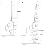
Phylogenetic reconstruction of the ORF2–3 overlapping region showed that the HEV isolated in Uruguay clustered in genotype 1 with high bootstrap values (Figure, panel A). The same was observed from the phylogenetic reconstruction of the ORF1 RdRp region (Figure, panel B). Furthermore, these analyses revealed a very close relationship of this strain isolated in Uruguay to a strain isolated in India, Yam 67 (99.5% nucleotide identity). Additionally, within the ORF1, the strain shares a high percentage of nucleotide identity (99%–100%) with the genotype 1 strains isolated in Cuba (Cub 11 and 13) and Venezuela (VNZ792), the 2 countries in Latin America in which genotype 1 has been reported to be associated with autochthonous cases. This relationship between these strains from South America and the Yam 67 strain from India warrants further investigation. In Uruguay, we have recently reported the full molecular characterization of a set of HEV strains isolated from patients with 9 sporadic cases in a 2-year period; all strains were genotype 3 (10). Here, we describe the detection and phylogenetic analysis of a locally-acquired indigenous case of HEV infection associated with genotype 1 in Uruguay. Although the detection of genotype 1 in 1/10 cases might have occurred by chance, this result supports an endemic circulation of HEV in Uruguay. Data presented here, together with recent advancements in molecular epidemiology of HEV infection in South America, reveal an epidemiologic picture more complex than we initially assumed.
Acknowledgments
We thank María del Carmen Viñoly and Programa de Desarrollo de las Ciencias Básicas for administrative support.
This work was partially supported by a PhD fellowship from Agencia Nacional de Innovación e Investigación (to S.M.).
References
- Kumar S, Subhadra S, Singh B, Panda BK. Hepatitis E virus: the current scenario. Int J Infect Dis. 2013;17:e228–33. DOIPubMedGoogle Scholar
- Herrera JL. Hepatitis E as a cause of acute non-A, non-B hepatitis. Arch Intern Med. 1993;153:773–5. DOIPubMedGoogle Scholar
- Hino K, Kondo T, Niwa H, Uchida T, Shikata T, Rikahisa T, A small epidemic of enterically transmitted non-A, non-B acute hepatitis. Gastroenterol Jpn. 1991;26:139–41 .PubMedGoogle Scholar
- Okamoto H, Takahashi M, Nishizawa T, Fukai K, Muramatsu U, Yoshikawa A. Analysis of the complete genome of indigenous swine hepatitis E virus isolated in Japan. Biochem Biophys Res Commun. 2001;289:929–36. DOIPubMedGoogle Scholar
- Yazaki Y, Mizuo H, Takahashi M, Nishizawa T, Sasaki N, Gotanda Y, Sporadic acute or fulminant hepatitis E in Hokkaido, Japan, may be foodborne, as suggested by the presence of hepatitis E virus in pig liver as food. J Gen Virol. 2003;84:2351–7. DOIPubMedGoogle Scholar
- Dalton HR, Hunter JG, Bendall RP, Hepatitis E. Curr Opin Infect Dis. 2013;26:471–8.PubMedGoogle Scholar
- Echevarría JM, González JE, Lewis-Ximenez LL, Lopes dos Santos DR, Munné MS, Pinto MA, Hepatitis E virus infection in Latin America: a review. J Med Virol. 2013;85:1037–45. DOIPubMedGoogle Scholar
- Inoue J, Takahashi M, Yazaki Y, Tsuda F, Okamoto H. Development and validation of an improved RT-PCR assay with nested universal primers for detection of hepatitis E virus strains with significant sequence divergence. J Virol Methods. 2006;137:325–33. DOIPubMedGoogle Scholar
- Zhai L, Dai X, Meng J. Hepatitis E virus genotyping based on full-length genome and partial genomic regions. Virus Res. 2006;120:57–69. DOIPubMedGoogle Scholar
- Mirazo S, Ramos N, Russi JC, Arbiza J. Genetic heterogeneity and subtyping of human Hepatitis E virus isolates from Uruguay. Virus Res. 2013;173:364–70. DOIPubMedGoogle Scholar
Figure
Cite This ArticleRelated Links
Table of Contents – Volume 20, Number 1—January 2014
| EID Search Options |
|---|
|
|
|
|
|
|

Please use the form below to submit correspondence to the authors or contact them at the following address:
Juan Arbiza, Iguá 4225, Postal Code 11400, Montevideo, UruguayJuan Arbiza, Iguá 4225, Postal Code 11400, Montevideo, Uruguay
Top