Volume 21, Number 1—January 2015
Dispatch
Acute Blastocystis-Associated Appendicular Peritonitis in a Child, Casablanca, Morocco
Emilie Fréalle1 , Dima El Safadi1, Amandine Cian, Estelle Aubry, Gabriela Certad, Marwan Osman, Agnès Wacrenier, Emmanuel Dutoit, Colette Creusy, François Dubos, and Eric Viscogliosi
Figure
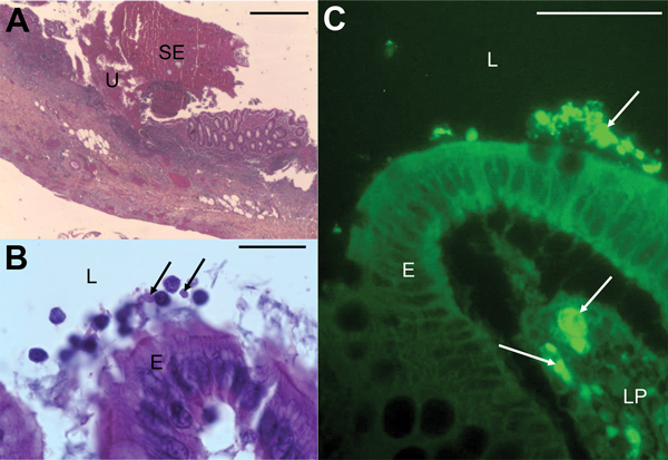
Figure. Micrographs showing histopathologic examination of appendix samples from a child who had peritonitis, Casablanca, Morocco, 2013. A) Ulceration (U) covered with suppurative and fibrinous exudates (SE) (hematoxylin-eosin stain). Scale bar indicates 200μm. B) Blastocystis parasites (arrows) in the lumen (L), and at the surface of the epithelium (E) (hematoxylin-eosin stain). Scale bar indicates 20μm. C) Blastocystis parasites (arrows) in the lumen, at the surface of the epithelium and in the lamina propria (LP) of the mucosa (immunofluorescence labeling with anti-Blastocystis ParaFlorB antibody). Scale bar indicates 50μm.
1These authors contributed equally to this article.
Page created: December 19, 2014
Page updated: December 19, 2014
Page reviewed: December 19, 2014
The conclusions, findings, and opinions expressed by authors contributing to this journal do not necessarily reflect the official position of the U.S. Department of Health and Human Services, the Public Health Service, the Centers for Disease Control and Prevention, or the authors' affiliated institutions. Use of trade names is for identification only and does not imply endorsement by any of the groups named above.